Le Fort fractures are complex fractures of the midface, involving the maxillary bone and surrounding structures in either a horizontal, pyramidal or transverse direction.
The hallmark of these fractures is traumatic pterygomaxillary separation. They account for between 10% and 20% of all facial fractures and can be potentially life-threatening and disfiguring.
Le Fort fractures were named after the French surgeon Rene Le Fort (1869-1951), who studied cadaver skulls that had been subjected to blunt force trauma. These experiments determined the areas of structural weakness of the maxilla, which were then specified as ‘lines of weakness’ where fractures can potentially occur.
Aetiology
The aetiology of Le Fort fractures varies according to the type of fracture. Common mechanisms include motor vehicle accidents, sporting injuries, assaults, and falls from a significant height. Patients with Le Fort fractures often have associated head and cervical spine injuries. They are also often other facial fractures, and neuromuscular injury and dental avulsions are common.
The type of fracture sustained depends upon the directionality of the force through the face:
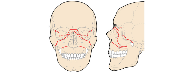
- Le Fort type I fractures – usually result from a force directed in a downward direction against the upper teeth
- Le Fort type II fractures – usually result from a force through the lower or mid maxilla
- Le Fort type III fractures – usually result caused by force through the nasal bridge and upper part of the maxilla.
The Le Fort classification system
The Le Fort classification system separates fractures of the midface into three different groups according to the plane of injury. As the classification increases, the anatomic level of the maxillary fracture ascends from inferior to superior with respect to the maxilla.
Le Fort I fractures
Le Fort I fractures are horizontal fractures across the inferior aspect of the maxilla. The teeth separate from the upper face, and the fracture extends through the lower nasal septum, the lateral maxillary sinus wall and into the palatine bones and pterygoid plates. These fractures are sometimes referred to as a ‘floating palate’, as they usually result in mobility of the hard palate from the midface. There is often accompanying facial oedema, loose teeth, dental fractures and malocclusion.
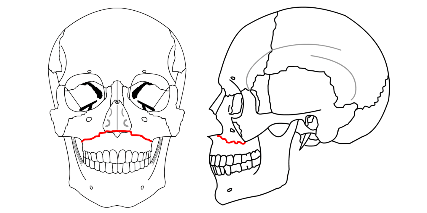
Le Fort I fracture, image sourced from Wikipedia
Courtesy of Rosario Van Tulpe CC BY-SA 3.0
Le Fort II fractures
Le Fort II fractures are pyramidal-shaped fractures, with the teeth at the base of the pyramid and the nasofrontal suture at the apex. The fracture line extends from the nasal bridge and passes through the superior wall of the maxilla, inferolaterally through the lacrimal bones and inferior orbital floor and rim, and inferiorly through the anterior wall of the maxillary sinus. These fractures are sometimes referred to as a ‘floating maxilla’, as they usually result in mobility of the maxilla from the midface. There is often accompanying facial oedema, epistaxis, subconjunctival haemorrhage, CSF rhinorrhoea, and telecanthus (widening and flattening of the nasal bridge).
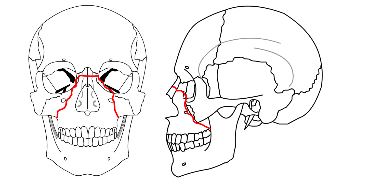
Le Fort II fracture, image sourced from Wikipedia
Courtesy of Rosario Van Tulpe CC BY-SA 3.0
Le Fort III fractures
Le Fort III fractures are transverse fractures of the midface. The fracture line passes the nasofrontal suture, the maxillofrontal suture, the orbital wall and the zygomatic arch and zygomaticofrontal suture. There is separation of all of the facial bones from the cranial base, and for this reason, they are sometimes referred to as craniofacial disjunction or ‘floating face’ fractures. These fractures are often accompanied by massive facial oedema, orbital ecchymoses and facial flattening, and the entire face can be shifted. They are the rarest and most serious of the three types of Le Fort fracture.
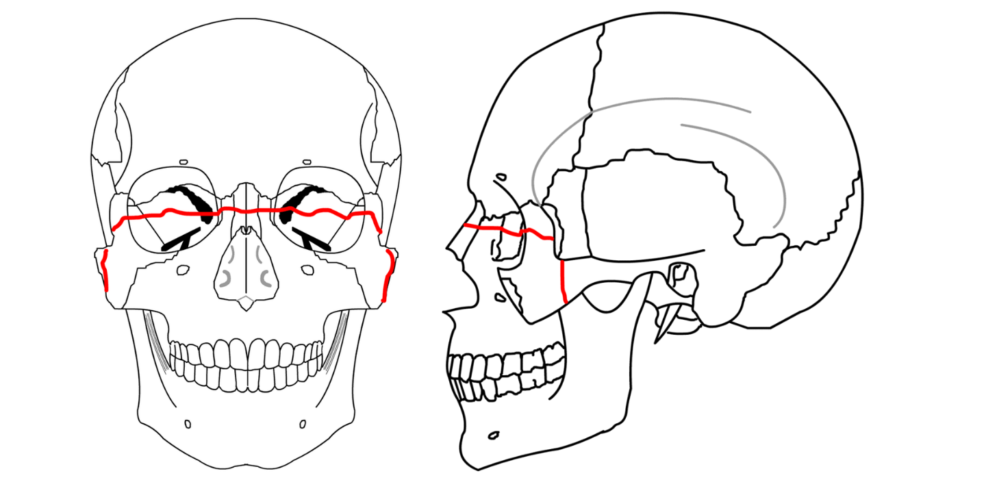
Le Fort III fracture, image sourced from Wikipedia
Courtesy of Rosario Van Tulpe CC BY-SA 3.0
A simple aide-mémoire
It can be challenging to memorise these three injury patterns; however, the following aide-mémoire can be helpful:
- Le Fort I – horizontal floating palate
- Le Fort II – pyramidal floating maxilla
- Le Fort III – transverse floating face
Management of Le Fort fractures
The patient should be evaluated and stabilised using the standard ATLS ‘ABCDE approach’. Once any life-threatening injuries have been addressed and the patient stabilised, the fractures are treated surgically.
All Le Fort fractures require fixation of unstable fracture segments to stable structures. The primary aims of surgical management are to:
- Restore the facial projection and the involved sinus cavities
- Re-establish proper occlusion of teeth; note that adequate repair cannot be performed without good occlusion
- Restore the integrity of the nose and orbit.
Header image used on licence from Shutterstock
Thank you to the joint editorial team of www.mrcemexamprep.net for this ‘Exam Tips’ blog post.