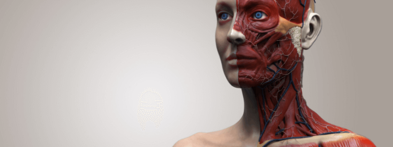
The triangles of the neck are an extremely common anatomy exam topic. In the first article, we covered the important knowledge requirements of the anterior triangle of the neck. In this second article, we focus on the posterior triangle of the nek.
The sternocleidomastoid muscle is the main muscular landmark of the neck and divides it diagonally into two triangular divisions:
- The anterior triangle of the neck
- The posterior triangle of the neck
These triangles are useful when communicating information about the location of structures and pathology in the neck, for example palpable masses and swellings in the clinical setting.
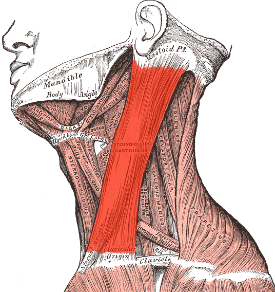
The sternocleidomastoid muscle (adapted from Gray’s Anatomy)
The posterior triangle of the neck
The boundaries of the posterior triangle of the neck are as follows:
- Anterior boundary – sternocleidomastoid muscle
- Posterior boundary – trapezius muscle
- Inferior boundary (base) – middle third of the clavicle
- Superior boundary (apex) – point that sternocleidomastoid and trapezius meet on the superior nuchal line of the occipital bone
- Roof – investing layer of cervical fascia
- Floor – muscles covered by the prevertebral layer of cervical fascia
The posterior triangle of the neck contains numerous important structures. It is easiest to consider these as nerves, vessels, and muscles separately.
The nerves contained within the posterior triangle of the neck are:
- The accessory nerve (CN XI)
- The phrenic nerve (C3,4,5)
- The suprascapular nerve
- Branches of the cervical plexus
- Roots and trunks of the brachial plexus
The vessels contained within the posterior triangle of the neck are:
- The 3rd part of the subclavian artery
- The transverse cervical artery
- The subscapular artery
- The occipital artery
- The terminal part of the external jugular vein
- The subclavian vein
- The brachiocephalic vein
The muscles contained within the posterior triangle of the neck are:
- The inferior belly of omohyoid muscle
- The anterior, middle, and posterior scalene muscles
- Levator scapulae muscle
- Splenius capitis muscle
The posterior triangle is subdivided into two further triangles by the omohyoid muscle:
- Occipital triangle
- Subclavian triangle
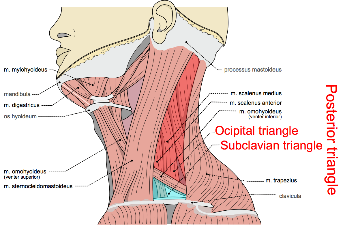
The posterior triangle of the neck and its subdivisions, image sourced from Wikipedia
Courtesy of Olek Remesz CC BY-SA 3.0
The occipital triangle
The occipital triangle is the larger, superior subdivision of the posterior triangle of the neck. It is formed by the following boundaries:
- Anterior boundary – sternocleidomastoid muscle
- Posterior boundary – trapezius muscle
- Inferior boundary – inferior belly of the omohyoid muscle
Its floor is formed by the splenius capitis, levator scapulae, and the middle and anterior scalene muscles. Its roof is formed by the superficial layer of the investing fascia
The accessory nerve (CN XI) crosses the upper half of the occipital triangle diagonally, passing from the deep surface of sternocleidomastoid inferiorly on levator scapulae to reach the deep surface of trapezius, innervating the sternocleidomastoid and trapezius muscles.
The subclavian triangle
The subclavian triangle, which is also known as the omoclavicular triangle, is the smaller, inferior subdivision of the posterior triangle of the neck. It is formed by the following boundaries:
- Anterior boundary – sternocleidomastoid muscle
- Superior boundary – inferior belly of the omohyoid muscle
- Inferior boundary – Clavicle
Its floor is formed by the splenius capitis, levator scapulae, and the middle and anterior scalene muscles. Its roof is formed by the superficial layer of the investing fascia.
For thousands of anatomy tutorials and questions visit: www.anatomyprep.co.uk