
The triangles of the neck are an extremely common anatomy exam topic. In this first of two articles, we will cover the important knowledge requirements of the anterior triangle of the neck.
The sternocleidomastoid muscle is the main muscular landmark of the neck and divides it diagonally into two triangular divisions:
- The anterior triangle of the neck
- The posterior triangle of the neck
These triangles are useful when communicating information about the location of structures and pathology in the neck, for example palpable masses and swellings in the clinical setting.
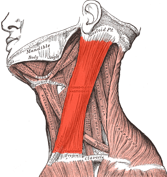
The sternocleidomastoid muscle (adapted from Gray’s Anatomy)
The anterior triangle of the neck
The boundaries of the anterior triangle of the neck are as follows:
- Anterior boundary – medial line of the neck
- Posterior boundary – sternocleidomastoid muscle
- Superior boundary (base) – inferior border of the mandible
- Inferior boundary (apex) – jugular notch at the superior border of the manubrium sterni
- Floor – formed by the pharynx, larynx, and thyroid gland
The anterior triangle is subdivided into four further triangles:
- Submental triangle
- Submandibular triangle
- Carotid triangle
- Muscular triangle
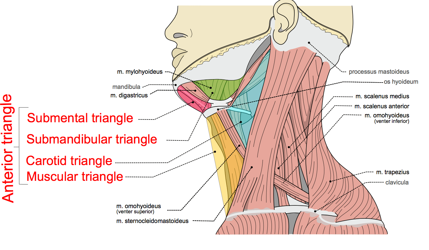
The anterior triangle of the neck and its subdivisions, image sourced from Wikipedia
Courtesy of Olek Remesz CC BY-SA 3.0
The submental triangle
The submental triangle is situated within the neck beneath the chin. It is formed by the following boundaries:
- Medial boundary – medial line of the neck
- Lateral boundary – anterior belly of the digastric muscle
- Inferior boundary – hyoid bone
The base of the submental triangle is formed by the mylohyoid muscle, which extends from the mandible to the hyoid bone.
The main contents of the submental triangle are:
- Submental lymph nodes
- Small veins that unite to form the anterior jugular vein
The submandibular triangle
The submandibular triangle is situated within the neck below the body of the mandible. It is formed by the following boundaries:
- Superior boundary – body of the mandible
- Anterior boundary – anterior belly of the digastric muscle
- Posterior boundary – posterior belly of the digastric muscle
The main contents of the submandibular triangle are:
- Submandibular gland
- Submandibular lymph nodes
- Hypoglossal nerve (CN XII)
- Mylohyoid nerve
- Parts of the facial artery and vein
The carotid triangle
The carotid triangle is situated medially to the upper part of the sternocleidomastoiud muscle. It is formed by the following boundaries:
- Superior boundary – posterior belly of the digastric muscle
- Lateral boundary – Medial border of the sternocleidomastoid muscle
- Inferior boundary – superior belly of the omohyoid muscle
The main contents of the carotid triangle are:
- Common carotid artery
- External carotid artery and some branches
- Internal carotid artery
- Hypoglossal nerve
- Vagus nerve
- Larynx and pharynx (deep)
- Internal laryngeal nerve (deep)
- External laryngeal nerve (deep)
The common carotid artery bifurcates within the carotid triangle to form the external and internal carotid arteries.
The muscular triangle
This muscular triangle actually has four sides and is situated more inferiorly than the other triangles. It is formed by the following boundaries:
- Superior boundary – hyoid bone
- Medial boundary – medial line of the neck
- Superolateral boundary – superior belly of the omohyoid muscle
- Inferolateral boundary – inferior portion of the sternocleidomastoid muscle
The main contents of the muscular triangle are:
- Sternothyroid muscle
- Sternohyoid muscle
- Thyroid gland
- Parathyroid glands
- Pharynx
- Trachea
- Oesophagus
Next: Triangles of the Neck Part 2 – The Posterior Triangle
For thousands of anatomy tutorials and questions visit: www.anatomyprep.co.uk


You make it so concise, point form, clear.