
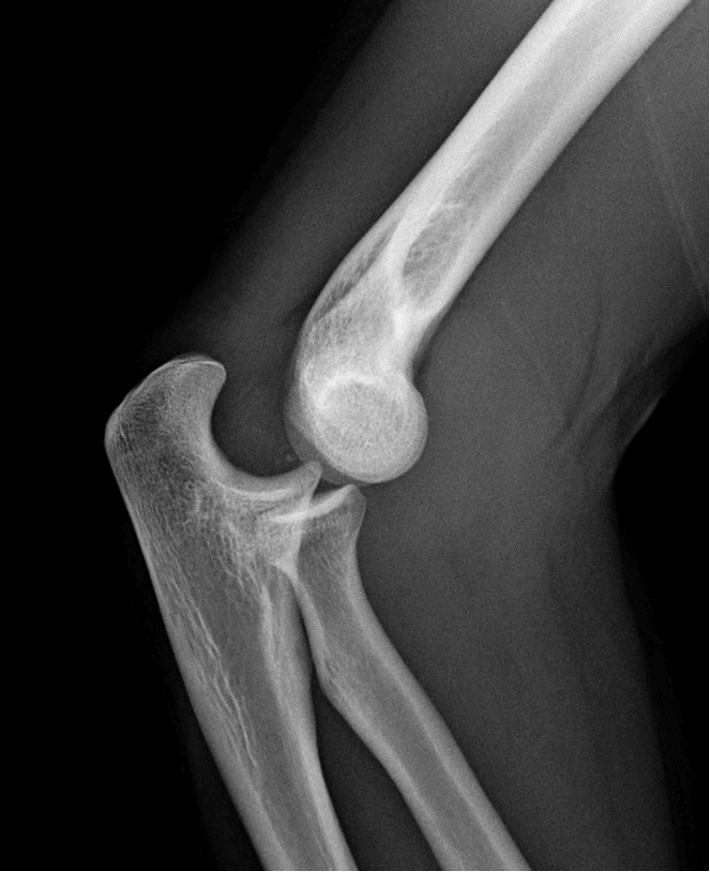
A 21-year-old football player presents with a deformed, severely painful elbow following a fall onto his right arm. An X-ray is performed, which is shown below.
Show Answer
This patient has a posterolateral elbow dislocation.
Elbow dislocations are the second commonest major joint dislocation after the shoulder in adults, and the most common joint dislocation in children. Approximately 80-90% of elbow dislocations are posterolateral.
Elbow X-ray sourced from Wikipedia
Courtesy of Dr. James Heilman CC BY-SA 3.0
Show Answer
Show Answer
Neurovascular injuries are relatively uncommon, but a thorough neurovascular examination should always be performed. The most commonly damaged structure is the ulnar nerve due to valgus stretching.
The median nerve can also be damaged, but this is less common. Damage to the brachial artery is rare and is typically associated with open dislocations. Damage to the brachial artery can be assessed for by palpating for a radial pulse. The most common associated fracture in complex dislocations is of the medial epicondyle.
Show Answer
The majority of elbow dislocations can be successfully relocated in the Emergency Department under conscious sedation. The options for reduction include the following two techniques:
- Flex the elbow to 60 degrees with counter-traction on the upper arm. Pull on the fully pronated forearm at this angle. Slight flexion at the elbow may be necessary.
- Lever the olecranon forward with both thumbs while holding the elbow flexed. An assistant then provides traction on the forearm.
Following a successful reduction, a repeat neurovascular examination should be performed, and the elbow should be immobilised in an above elbow backslab POP at 90 degrees. A repeat X-ray should be performed. Some patients require admission for elevation of the arm and observation for significant limb swelling.
Header image used on licence from Shutterstock


The article is useful
The article is useful and practical