The cell is the smallest living thing in the human organism, it is the functional and structural unit of all living things and all living structures in the human body are made of cells.
The human body contains several billion cells, encompassing a vast range of types with hundreds of specialised functions. Cells differ widely in shape—round, flat, long and thin, or short and thick—as well as in size. For example, cerebellar granule cells in the brain are among the smallest at around 4 micrometres, while oocytes (egg cells) in the female reproductive system can reach up to 100 micrometres.
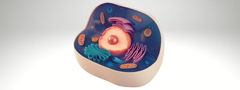
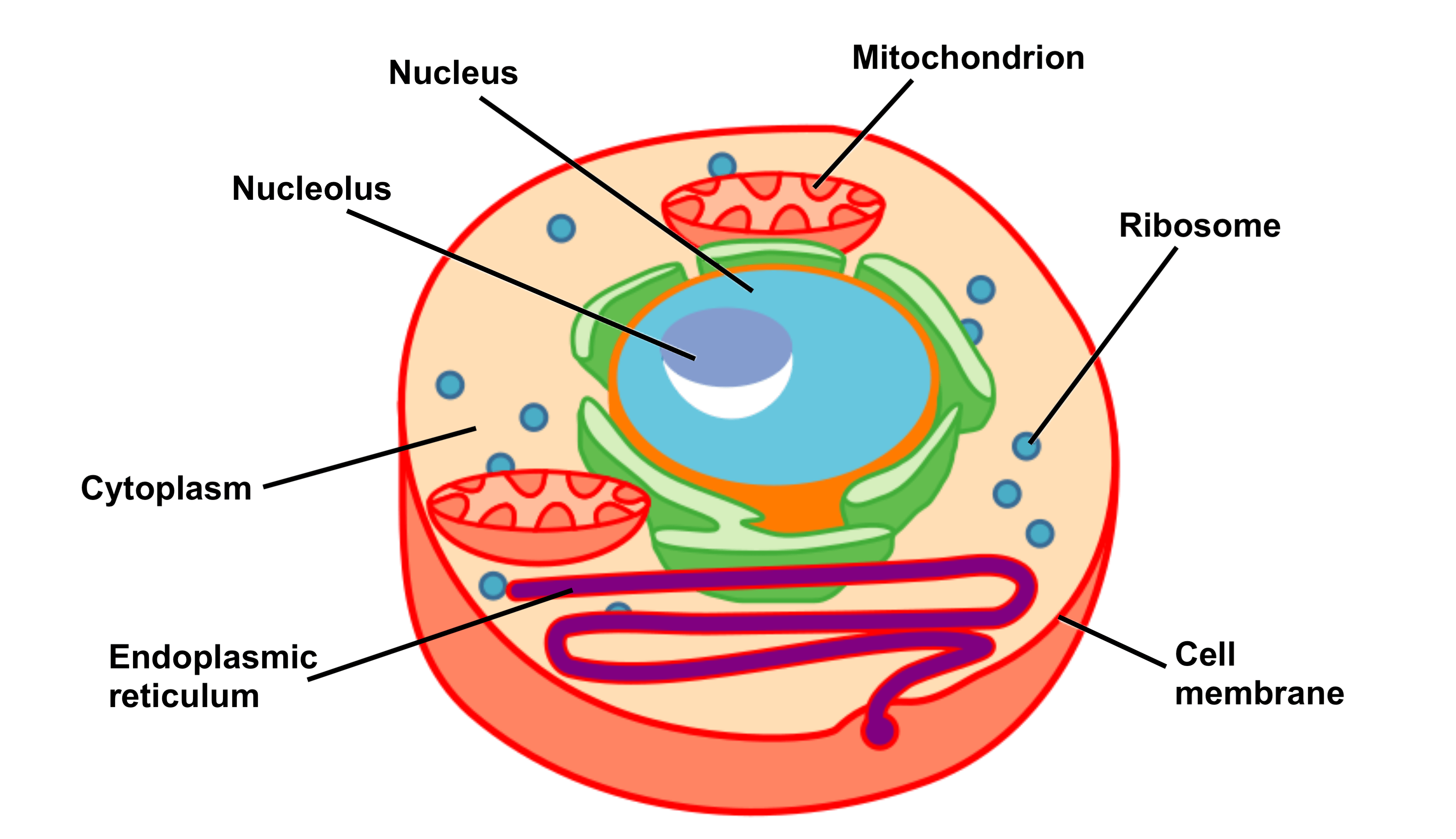
A labelled diagram of a human cell is shown below. Each of the labelled structures will then be discussed individually.
Cytoplasm
The cytoplasm is a jelly-like substance that makes up most of the cell’s volume and is composed primarily of water (around 90%). It also contains dissolved nutrients, salts, and waste products.
Its main role is to support and suspend the organelles within the cell. Additionally, it supplies essential salts and sugars for nourishment and serves as the medium in which metabolic reactions take place.
The main functions of the cytoplasm are:
- It provides mechanical support to the cell and keep the shape of the cell, which is achieved by exerting pressure against the cell membrane (turgor pressure).
- It acts a storage area for small carbohydrate, lipid and protein molecules.
- It nourishes the cell by supplying it with salts and sugars and provides a medium for metabolic reactions to occur in.
- It is the site of most cellular activities including metabolism and cell division.
- It contains ribosomes which are essential for protein synthesis.
The nucleus
The nucleus is the largest organelle within the cell and houses the majority of its genetic material. It is surrounded by a double lipid membrane known as the nuclear envelope, which separates the nucleus from the surrounding cytoplasm.
Embedded within this envelope are small openings called nuclear pores, which regulate the exchange of materials—such as proteins and RNA—between the nucleus and the cytoplasm.
The nucleus also contains the nucleolus, a smaller structure that lacks a surrounding membrane and occupies approximately 25% of the volume of the nucleus. The primary function of the nucleolus is to transcribe ribosomal RNA (rRNA) and combine it with proteins to form incomplete ribosomes. rRNA is important for the construction of ribosomes, which are the site of protein translation.
Other functions of the nucleus include:
- The control gene expression and facilitate the replication of DNA during the cell cycle.
- The production of messenger RNA (mRNA) which encodes for enzymes and, therefore, assists with the control of the metabolic functions of cells.
- The control of the structure of the cell via the transcription of DNA which encodes for structural proteins.
Mitochondria
Mitochondria are membrane-bound organelles responsible for producing the cell’s chemical energy. They do this by using oxygen to break down sugars and small fatty acids, generating adenosine triphosphate (ATP)—the cell’s main energy-carrying molecule. This energy production process is called oxidative phosphorylation and relies on the enzyme ATP synthase. ATP is then released when needed to power various cellular activities.
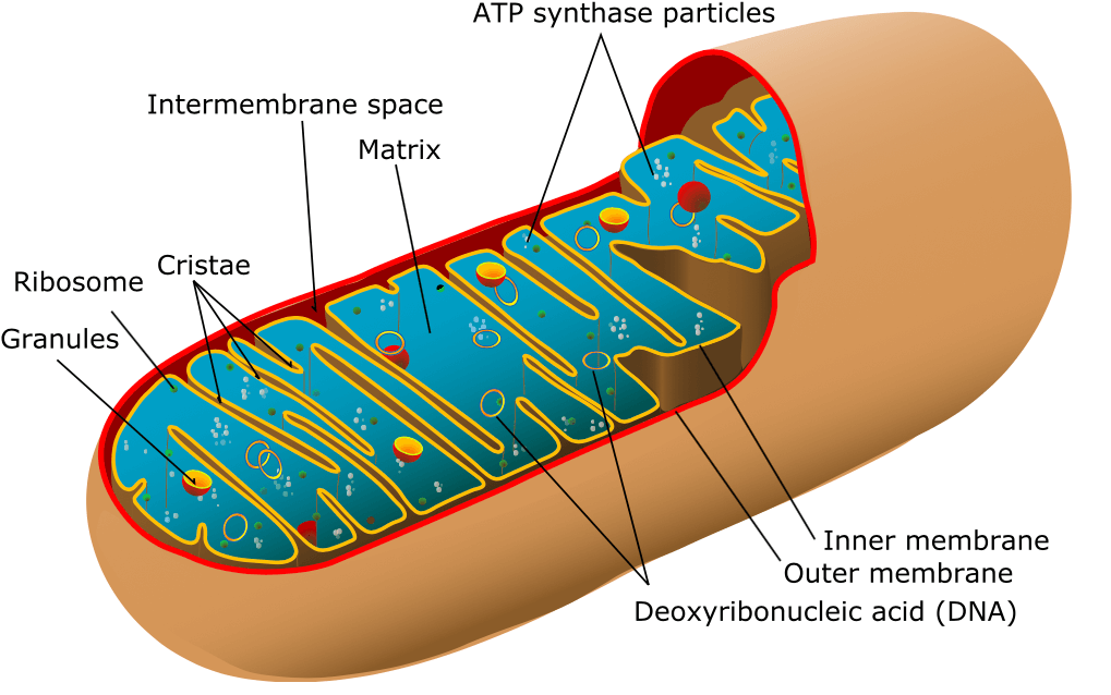
Mitochondria have two phospholipid bilayers: an outer membrane and a highly folded inner membrane. The folds of the inner membrane, known as cristae, increase surface area and contain specialised proteins essential for ATP production. Between these membranes is the intermembrane space, which stores proteins involved in cellular respiration. Inside the inner membrane lies the mitochondrial matrix—a jelly-like substance rich in ATP synthase and other enzymes critical for energy metabolism.
A labelled diagram of a mitochondrion is shown below:
Endoplasmic reticulum
The endoplasmic reticulum (ER) is the site where proteins are assembled by ribosomes. Located in the cytoplasm, the ER consists of a double membrane shaped into a network of hollow tubes, flattened sheets, and rounded sacs known collectively as cisternae. It is also directly connected to the nuclear envelope.
There are two types of ER: smooth and rough.
-
Smooth ER lacks ribosomes on its surface, giving it a smooth appearance. It is involved in lipid synthesis—including oils, phospholipids, and steroids—as well as carbohydrate metabolism, calcium regulation, and drug detoxification.
-
Rough ER is studded with ribosomes, giving it a rough appearance. It plays a key role in protein synthesis and also contributes to membrane production. The folds in its membrane increase surface area, allowing more ribosomes to attach and boosting protein output.
A 3D representation of the ER is shown below:
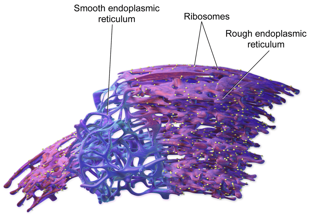
Image sourced from Wikipedia
Courtesy of Medical Gallery of Blausen Medical CC BY-SA 3.0
Ribosomes
Ribosomes are composed of RNA and protein and play an important role in protein synthesis. They are found in the cytoplasm and can occur alone, in groups or attached to the endoplasmic reticulum, forming the rough ER (see above).
Ribosomes bind messenger RNA (mRNA) to form a structure known as a polyribosome. They then use the information contained within the mRNA to build a protein with a specific amino acid sequence in a process is called translation.
The Golgi apparatus
The Golgi apparatus is responsible for the post-translational processing of newly made proteins. It consists of a stack of flat membrane-bound sacs called cisternae and is found in close proximity to the nucleus and endoplasmic reticulum.
Proteins are transported from the rough ER to the Golgi apparatus, where they are modified and packaged into vesicles. The Golgi apparatus then transfers these proteins to another location within the cell where they are required. For this reason, the Golgi apparatus is sometimes referred to as the ‘post office’ of the cell.
Vesicles and lysosomes
Vesicles are small, membrane-bound spherical sacs involved in the metabolism, transport, and storage of molecules within the cell. They are formed by both the Golgi apparatus and the endoplasmic reticulum, and can also bud off from the cell membrane. Vesicles are classified based on their contents and function—for example, transport vesicles move molecules between different parts of the cell.
Lysosomes are a specific type of vesicle produced by the Golgi apparatus or endoplasmic reticulum. They contain powerful enzymes capable of breaking down cellular components and food molecules such as carbohydrates and proteins. Lysosomes are especially abundant in animal cells that take in food via vacuoles. When a cell dies, lysosomes release their enzymes to break down and recycle the cell’s components.
Next: Cell Structure and Function Part 2– The Cell Membrane
Thank you to the joint editorial team of www.mrcemexamprep.net for this article.


Wonderful resource.
Thanks
I have appreciated this source coz of its detailed information, please may you continue servicing the world 🌎👏