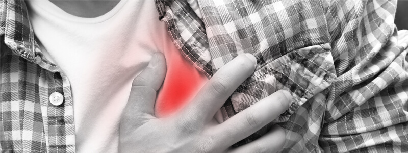
A 37-year-old man is involved in a high-speed motor vehicle collision. He was not wearing a seat belt and struck his chest on the steering wheel. He has presented complaining of severe central chest pain and breathlessness. You are the trauma call team leader. His observations are as follows: HR 110, BP 80/51, SaO2 95% on air, GCS 15/15. On examination he has extensive bruising over the centre of his chest, his neck veins are clearly distended and on auscultation his heart sounds are muffled.
1. Which potentially life-threatening chest injury has occurred in this case?
Show Answer
This patient has developed cardiac tamponade. This most commonly occurs from penetrating injuries but can also occur following blunt thoracic trauma. The trauma causes the pericardial cavity to fill with blood from the heart, great vessels, or pericardial vessels.
The pericardial cavity (also referred to as the pericardial space), contains the lubricating pericardial fluid, and is found between the parietal and visceral pericardium. The pericardial cavity usually contains around 20 ml of pericardial fluid. Once around 200 ml of fluid has accumulated systolic dysfunction and impairment of cardiac output begin to develop. This is referred to as cardiac tamponade.
Cardiac tamponade can be recognised clinically by Beck’s triad of:
- Distended neck veins
- Muffled heart sounds
- Hypotension
2. Other than trauma, what else can cause this condition?
Show Answer
Cardiac tamponade commonly occurs as a result of trauma but has numerous other causes including:
- Myocardial rupture
- Cancer
- Uraemia
- Pericarditis
- Cardiac surgery
3. How should this patient be treated?
Show Answer
This patient needs an urgent pericardiocentesis. Pericardiocentesis is the process by which excess fluid can be drained from the pericardial cavity.
The procedure can be performed blind or under guidance by CT or ultrasound scan. The current consensus opinion is that it should only be carried out blindly in life-threatening circumstances.
The standard approach is the subxiphoid approach, with the clinician standing to the patient’s right. The patient should be sat up at a 30-45 degree angle, which allows the pericardial fluid to pool inferiorly.
The left xiphocostal angle should be located and the needle introduced about 1 cm below this point. The needle should be introduced at approximately a 30 degree angle and aimed at the midpoint of the left clavicle. A give is sometimes felt as the needle pierces the parietal pleura and enters the pericardial cavity. At this point a flashback of fluid should be seen, which confirms correct needle placement.
Header image used on licence from Shutterstock


