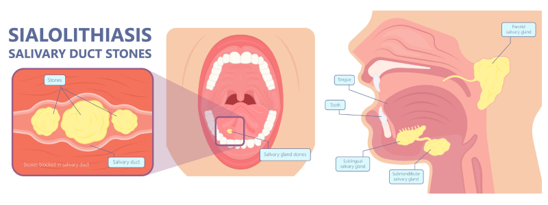
Sialolithiasis is a condition where a calcified stone (sialolith) forms within a salivary gland.
The salivary glands
The salivary glands are exocrine glands that produce saliva and secrete it into the oral cavity. There are three main salivary glands and several smaller ones of less significance.
The three main salivary glands are:
- The parotid glands
- The submandibular glands
- The sublingual glands
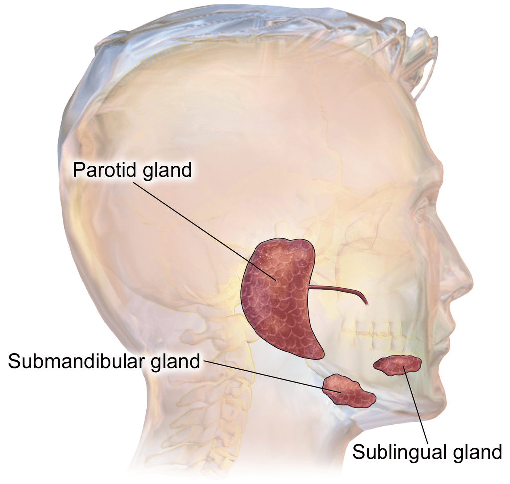
The salivary glands, image sourced from Wikipedia
Courtesy of Medical gallery of Blausen Medical CC BY-SA 3.0
Approximately 90% of salivary gland stones occur in the submandibular gland (Wharton’s duct), with the majority of the remainder occurring within the parotid gland. Occasionally they can also occur in the sublingual gland or minor salivary glands.
Risk factors for stone formation
There are several risk factors for the formation of salivary gland stones. These include:
- Dehydration
- Smoking
- Local trauma
- Sjogren’s syndrome
- History of nephrolithiasis
- Hyperparathyroidism
- Chronic periodontal disease
- Gout
- Chronic periodontal disease
- Head and neck radiotherapy
- Drugs, e.g. diuretics and anti-cholinergic medications
Clinical features
Sialolithiasis is commonly asymptomatic; however, a small percentage of people become symptomatic and experience intermittent symptoms. The sialolith obstructs the passage of saliva, which can cause pain and swelling of the affected gland during eating.
Pain is typically at its worst when salivary flow is high, immediately before and during eating, and then slowly subsides in the hour after. Symptoms are typically unilateral in nature.
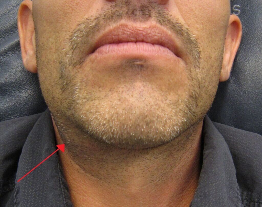
Swelling of the right submandibular gland due to an obstructing stoneWikipedia
Courtesy of Dr. James Heilman CC BY-SA 3.0
Bimanual palpation of the floor of the mouth may reveal a stone, and occasionally, the stone may actually be visible intra-orally at the duct orifice. If superimposed infection is present, it may be possible to express pus from the gland.

Visible stone seen in the submandibular duct Wikipedia
Courtesy of Dr. James Heilman CC BY-SA 3.0
Investigations
Acidic foods, such as lemon juice, can be used to stimulate salivary flow and may promote spontaneous expulsion of the stone.
Most suspected cases are investigated with either:
- X-rays of the floor of the mouth, which can demonstrate the stone as around 80% are radio-opaque, or
- Ultrasound scans – minimally invasive, cheap and very good at analysing the glands and peri-glandular structures, or
- Sialography – but this is not routinely performed because it is invasive compared to the other tests as it requires the gland to be cannulated and injected with dye.
Management
Most cases can be managed conservatively with rehydration, analgesia and the use of sialagogues, such as lemon juice, which can help to promote saliva production. In cases where the gland becomes infected, and sialadenitis develops, antibiotics are usually required.
Recurrent or persistent cases require definitive treatment with either interventional radiological procedures or surgery.
Header image used on license from Shutterstock


