A. Stomach
B. Liver
C. Spleen
D. Pancreas
E. Small intestine
Show Answer
The peritoneal cavity can be divided into the greater and lesser peritoneal sacs. The greater sac comprises the majority of the peritoneal cavity. The lesser sac is smaller and lies posterior to the stomach and lesser omentum.
The greater sac can be further subdivided into two compartments by the mesentery of the transverse colon (the transverse mesocolon):
- The supracolic compartment – which lies above the transverse mesocolon
- The infracolic compartment – which lies below the transverse mesocolon
The infracolic compartment is further subdivided into the left and right infracolic spaces by the mesentery of the small intestine.
The contents of the supracolic and infracolic compartments is shown in the table below:
| Supracolic compartment | Infracolic compartment |
|---|---|
| Stomach Liver Spleen | Small intestine Ascending colon Descending colon |
A. Left gastric artery
B. Right gastric artery
C. Short gastric arteries
D. Left gastro-omental artery
E. Right gastro-omental artery
Show Answer
The stomach receives an extensive arterial supply that is derived from the coeliac trunk and its branches. Anastomoses form along the lesser curvature by the left and right gastric arteries and long the greater curvature by the left and right gastro-omental arteries.
The arteries supplying the stomach are:
- The left gastric artery – arises directly from the coeliac trunk
- The right gastric artery – is a branch of the common hepatic artery, which arises from the coeliac trunk
- The left gastro-omental artery – is a branch of the splenic artery, which arises from the common hepatic artery
- The right gastro-omental artery – is a terminal branch of the gastroduodenal artery, which arises from the common hepatic artery
- Short gastric arteries – arise from the distal end of the splenic artery
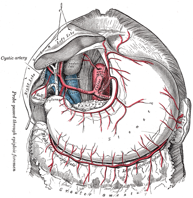
The arterial supply of the stomach (from Gray’s Anatomy)
A. Duodenum
B. Ileum
C. Jejenum
D. Caecum
E. Ascending colon
Show Answer
A Meckel’s diverticulum is a vestigial remnant of the vitellointestinal duct. It is the commonest malformation of the gastrointestinal tract, being present in around 2% of the population. They are twice as common in men than women.
When present a Meckel’s diverticulum is located in the distal ileum, usually within 60-100 cm (2 feet) of the ileocaecal valve. They are usually 3-6 cm (approx. 2 inches) long and may have a greater lumen than that of the ileum.
They are commonly found as an incidental finding, particularly at appendicectomy. The majority are asymptomatic but they can present with the following complications:
- Haemorrhage (25-50% of complications)
- Intestinal obstruction (10-40% of complications)
- Diverticulitis
- Perforation
Meckel’s diverticula run antimesenterically but receive their blood supply from the mesentery of the ileum, and a typical feeding vessel called the vitelline artery can be identified. They are typically lined with ileal mucosa but frequently contain ectopic mucosa, the two commonest types being gastric (50%) and pancreatic (5%). More rarely colonic or jejunal mucosa may be present.
The ‘rule of 2s’ is a useful aid-mémoire
- 2% of the population
- 2:1 male:female ratio
- 2 feet from the ileocaecal valve
- 2 inches in length
- 2 types of common ectopic tissue (gastric and pancreatic)
- 2 years most common age at clinical presentation
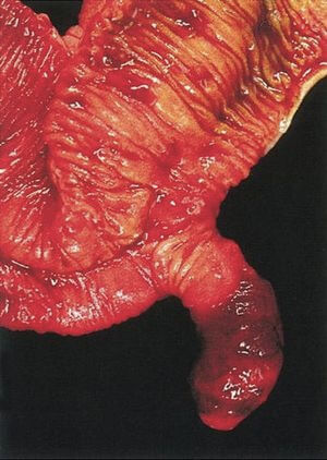
Surgical specimen of a Meckel’s diverticulum
A. One-third of the distance from the anterior superior iliac spine to the umbilicus
B. One-third of the distance from the posterior superior iliac spine to the umbilicus
C. Two-thirds of the distance from the anterior superior iliac spine to the umbilicus
D. Two-thirds of the distance from the posterior superior iliac spine to the umbilicus
E. Midway between the anterior superior iliac spine and the umbilicus
Show Answer
Appendicitis is acute inflammation of the appendix and is one of the most common causes of the acute abdomen. It is typically a disease of children and young adults with a peak incidence in the 2nd to 3rd decades of life.
The classical presentation of appendicitis is with poorly localized periumbilical pain (referred pain from the visceral peritoneum). Within a day or two this pain typically localizes to McBurney’s point (pain from parietal peritoneum). There is usually associated fever, anorexia and nausea.
McBurney’s point is defined as being the point that lies one-third of the distance from the anterior superior iliac spine to the umbilicus. It roughly corresponds with the most common position of the attachment of the base of the appendix to the caecum.
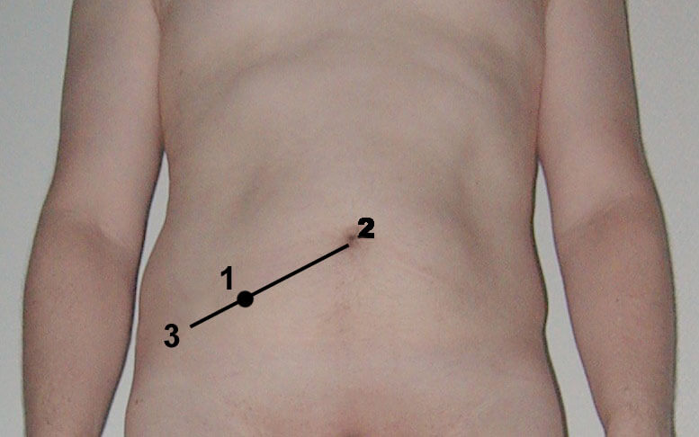
Image sourced from Wikipedia
Courtesy of Steven Fruitsmaak CC BY-SA 3.0
A. L3
B. T11
C. T12
D. L1
E. L2
Show Answer
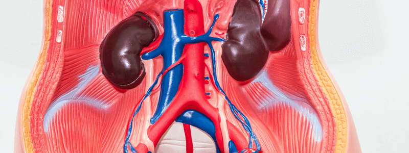
The abdominal aorta is the largest artery in the abdominal cavity. It arises as a continuation of the thoracic aorta as it passes posterior to the median arcuate ligament of the diaphragm at the T12 vertebral level.
It descends caudally in the retroperitoneal space, anterior and to the left of the lumbar vertebral bodies and to the left of the inferior vena cava.
It is approximately 13 cm long and ends slightly to the left of the midline at the L4 vertebral level where it bifurcates into the right and left common iliac arteries.
The abdominal aorta has three main groups of branches:
- Single ventral gut arteries:
- Coeliac artery (T12) – supplies the stomach, abdominal oeseophagus, liver, spleen, superior pancreas, and superior duodenum
- Superior mesenteric artery (L1) – supplies the distal duodenum, jejunum. Ileum, ascending colon and part of the transverse colon
- Inferior mesenteric artery (L3) – supplies the large intestine from the splenic flexure to the upper part of the rectum
- Paired visceral arteries:
- Middle adrenal arteries (L1) – supply the adrenal glands
- Renal arteries (L2) – supply the kidneys
- Gonadal arteries (L2/L3) – supply the testicles/ovaries
- Paired wall arteries:
- Inferior phrenic arteries (T12) – supply the diaphragm
- Four paired lumbar arteries (between L1 and L4) – supply the abdominal wall and spinal cord
It also gives off one single unpaired parietal artery, the median sacral artery, at the L4 vertebral level.
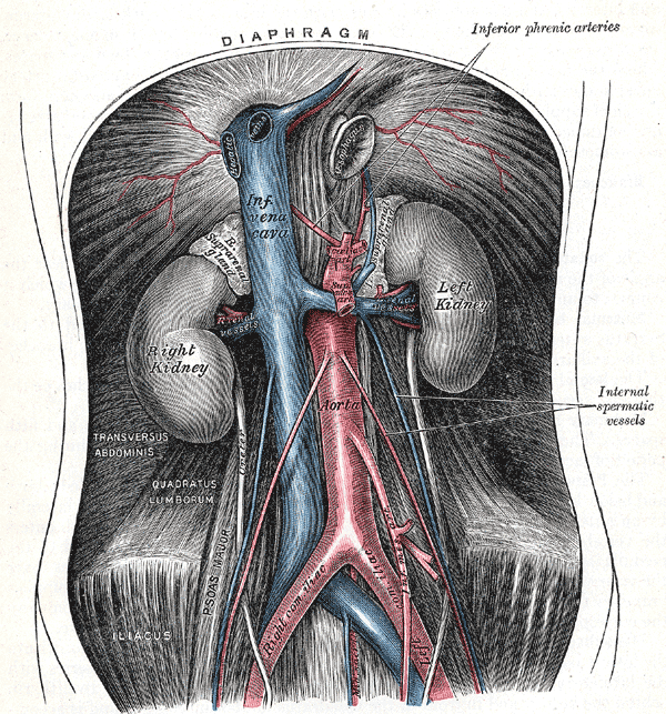
The abdominal aortic, its relations and branches (from Gray’s Anatomy)
For thousands more anatomy tutorials and questions visit: www.anatomyprep.co.uk

Header image used on licence from Shutterstock.


Nice topic
This is very nice and also useful.
excellent review
Nice review