Test your knowledge of lower limb anatomy with these questions.
1. Which SINGLE statement is true regarding the blood supply to the lower limb?
A. The femoral artery arises directly from the internal iliac artery
B. The profunda femoris arises from the medial side of the femoral artery
C. The fibular (peroneal) artery is a branch of the anterior tibial artery
D. The popliteal artery begins in Hunter’s canal
E. The dorsalis pedis artery is a direct continuation of the posterior tibial artery
Show Answer
Answer: D. The popliteal artery begins in Hunter’s canal
The popliteal artery begins as a continuation of the femoral artery as it traverses Hunter’s canal (the adductor hiatus).
The femoral artery arises from the external iliac artery.
The profunda femoris arises from the lateral side of the femoral artery within the femoral triangle.
The dorsalis pedis is a direct continuation of the anterior tibial artery distal to the ankle joint.
The fibular (peroneal) artery is a branch of the posterior tibial artery.
2. Which of the following is the most medial of the contents of the femoral triangle?
A. Femoral artery
B. Femoral nerve
C. Femoral canal
D. Femoral vein
E. Sartorius muscle
Show Answer
Answer: C. Femoral canal
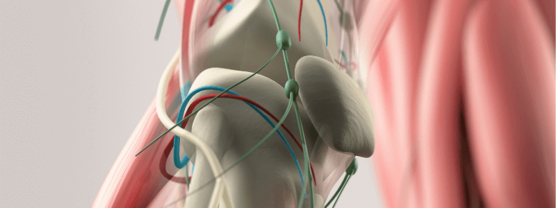
The femoral triangle is an anatomical region situated in the upper thigh area. It appears as a triangular depression that is situated inferior to the inguinal ligament when the thigh is flexed, abducted and laterally rotated.
The borders of the femoral triangle can be remembered by the mnemonic ‘SAIL’ (The femoral triangle is shaped like the sail of a ship):
- Laterally – Medial border of Sartorius
- Medially – Medial border of Adductor longus
- Superiorly – Inguinal Ligament
Its floor is formed by pectineus and adductor longus medially and iliopsoas laterally. Its roof is formed by the fascia lata.
The following structures are contained within the femoral triangle from lateral to medial:
- Femoral nerve
- Femoral artery
- Femoral vein
- Femoral canal (contains deep inguinal lymph nodes and lymphatic vessels)
The femoral artery, vein and canal are contained with a fascial compartment known as the femoral sheath.
The femoral triangle is clinically important as it provides easy access to the femoral artery, which can be utilised for angioplasty and central venous cannulation. It also provides easy access to the femoral nerve, which can be utilised for femoral nerve blocks.
The femoral triangle and its contents (from Gray’s Anatomy).
3. The great saphenous vein passes posteriorly to which of the following landmarks?
A. Lateral malleolus of the ankle
B. Medial malleolus of the ankle
C. Calcaneal tendon
D. Medial epicondyle of the femur
E. Lateral epicondyle of the femur
Show Answer
Answer: D. Medial epicondyle of the femur
The great saphenous vein is a large, subcutaneous, superficial vein of the leg. It is the longest vein in the body, running the entire length of the lower limb.
It is formed by the dorsal venous arch in the foot in combination with the dorsal vein of the great toe. It then ascends up the medial side of the leg, passing anteriorly to the medial malleolus at the ankle.
At the knee it runs over the posterior border of the medial epicondyle of the femur. It then courses anteriorly to lie on the anterior surface of the thigh before entering the saphenous opening in the fascia lata.
The great saphenous vein terminates by draining into the femoral vein immediately inferior to the inguinal ligament.
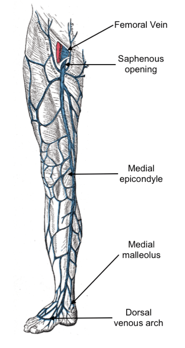
The surface markings of the great saphenous vein (adapted from Gray’s Anatomy)
4. Which of the following muscles is supplied by a branch of the posterior division of the femoral nerve?
A. Pectineus
B. Rectus femoris
C. Iliacus
D. Sartorius
E. Psoas major
Show Answer
Answer: B. Rectus femoris
The muscles supplied by the femoral nerve and its two divisions are shown in the table below:
| Branch | Muscles supplied |
|---|---|
| Femoral nerve in the abdomen | Iliacus |
| Anterior division of the femoral nerve | Pectineus Sartorius |
| Posterior division of the femoral nerve | Rectus femoris Vastus lateralis Vastus intermedius Vastus medialis |
5. Which of the following best describes the sensory area supplied by the lateral femoral cutaneous nerve
A. The lateral portion of the thigh
B. The central and medial portions of the thigh
C. The posterior aspect of the thigh
D. The anterior and lateral surface of the calf
E. The lateral surface of the calf
Show Answer
Answer: A. The lateral portion of the thigh
The lateral femoral cutaneous nerve (also known as lateral cutaneous nerve of the thigh) is a direct branch from the lumbar plexus that arises from the dorsal divisions of lumbar nerves L2 and L3. It is a purely sensory nerve innervates the skin over the lateral aspect of the thigh.
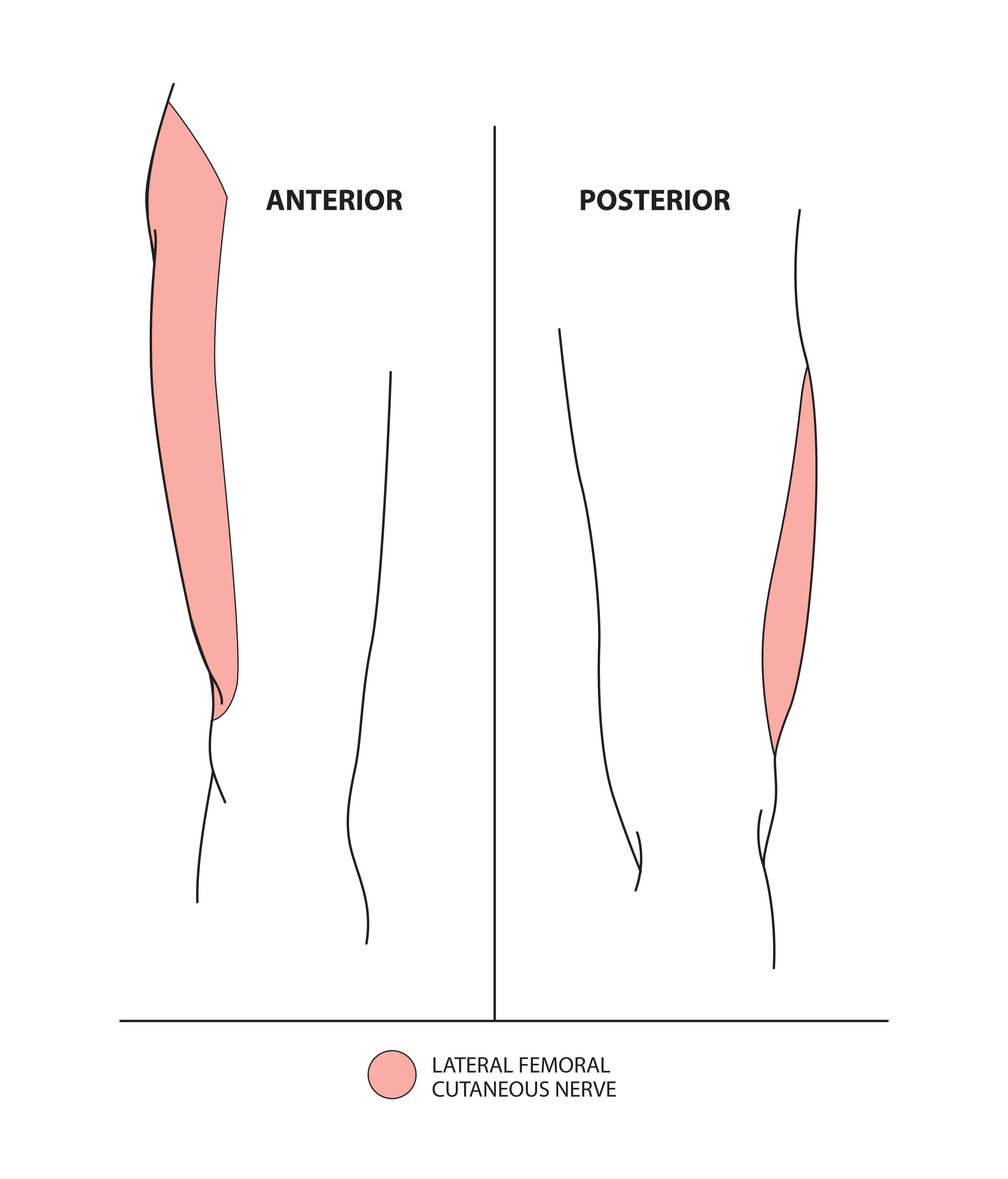
The sensory area supplied by the lateral femoral cutaneous nerve © Medical Exam Prep
For thousands more anatomy tutorials and questions visit: www.anatomyprep.co.uk

Header image used on licence from Shutterstock.


Good
V.nice
👌 perfect