1. The body of the sternum extends across which vertebral levels?
A. T2 to T4
B. T3 to T7
C. T4 to T6
D. T5 to T9
E. T6 to T11
Show Answer
The sternum is the flat, elongated bone that forms the middle of the anterior part of the thoracic cage. It connects to the ribs via cartilage, forming the front of the rib cage, and helps to protect the heart, great vessels, and lungs from injury.
It consists of three distinct parts:
- The manubrium: this is triangular shaped and lies at the T3/4 vertebral level. The superior border is indented by the suprasternal notch. Two clavicular notches lie on either side that articulate with the medial end of the clavicle. The 1st rib fuses with the lateral border just inferior to this.
- The body: this is longer, narrower, and thinner than the manubrium and extends from the T5 to T9 vertebral levels. The body’s nearly flat anterior surface is marked by three transverse ridges in the adult that represent the lines of fusion of its four originally separate segments.
- The xiphoid process: this is the smallest and most variable part of the sternum. It is cartilaginous in young people but is usually fully ossified by 40 years of age.
The body connects with the manubrium via the manubriosternal joint. Here the manubrium and the body lie in slightly different planes, forming the sternal angle, which lies at the T4/5 vertebral level. This readily palpable clinical landmark is located opposite the 2nd pair of costal cartilages.
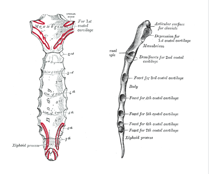
The sternum (from Gray’s Anatomy)
A. Internal carotid artery
B. External carotid artery
C. Axillary artery
D. Brachial artery
E. Subclavian artery
Show Answer
The superior thoracic aperture (also referred to as the thoracic inlet) is the opening at the top of the thoracic cavity.
The superior thoracic aperture is bounded by:
- Anteriorly: the superior border of the manubrium sterni and the costal cartilage of the first rib
- Laterally: the first pair of ribs
- Posteriorly: the first thoracic vertebra (T1)
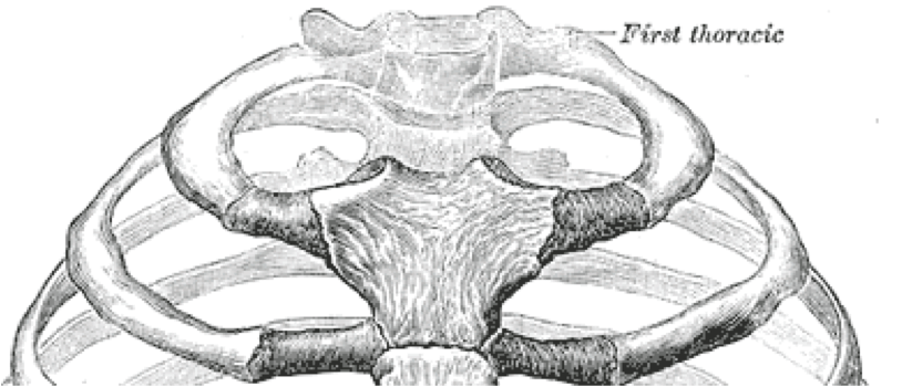
The superior thoracic aperture (from Gray’s Anatomy)
Several important structures pass through the superior thoracic aperture:
- The trachea
- The oesophagus
- The thoracic duct
- The apexes of the lungs
- Nerves (phrenic nerve, vagus nerve, recurrent laryngeal nerves, sympathetic trunks)
- Arteries (common carotid arteries, subclavian arteries)
- Veins (internal jugular veins, brachiocephalic veins, subclavian veins)
- Lymph nodes and lymphatic vessels
A. The oesophagus
B. Branches of the right phrenic nerve
C. Oesophageal branches of left gastric vessels
D. The vagal trunks
E. The thoracic duct
Show Answer
The diaphragm has three main openings:
- The caval opening: passes through the central tendon
- The oesophageal hiatus: situated in the posterior part of the diaphragm, to the left of the central tendon through the muscular sling of the right crus
- The aortic hiatus: situated in the posterior part of the diaphragm, between the left and right crus
The main openings in the diaphragm and their contents are shown in the table below:
| Opening | Vertebral Level | Contents |
|---|---|---|
| Caval opening | T8 | Inferior vena cava Branches of right phrenic nerve |
| Oesophageal hiatus | T10 | Oesophagus Anterior vagal trunk Posterior vagal trunk Oesophageal branches of left gastric vessels |
| Aortic hiatus | T12 | Aorta Azygous veins Thoracic duct |
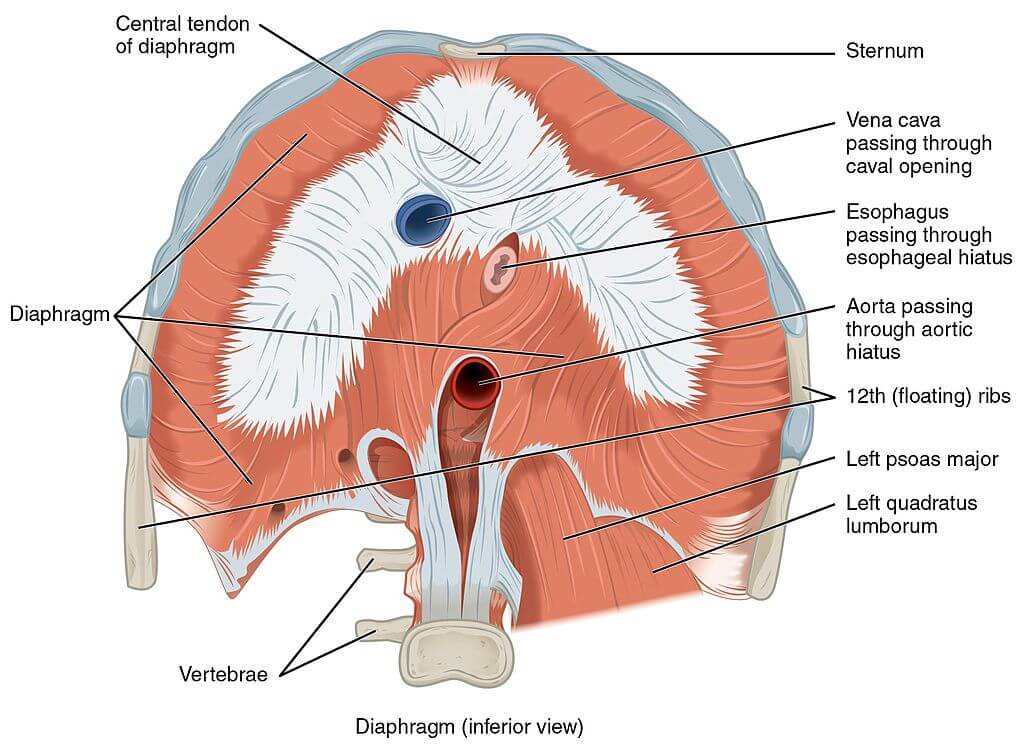
Image sourced from Wikipedia
Courtesy of OpenStax College CC BY-SA 3.0
4. Which SINGLE statement regarding the bronchi is FALSE?
A. The carina lies approximately at the level of the sternal angle
B. The left main bronchus is longer than the right
C. The left main bronchus is thinner than the right
D. The right main bronchus enters the right lung opposite the 5th thoracic vertebra
E. The left main bronchus is more vertical than the right
Show Answer
The left main bronchus is thinner but longer than the right. It is typically 5 cm long and enters the left lung opposite the 6th thoracic vertebra. It passes beneath the arch of the aorta and crosses in front of the oesophagus, the thoracic duct and the descending aorta.
The right main bronchus is wider, shorter and more vertical than the left. It is typically 2.5 cm long and enters the right lung opposite the 5th thoracic vertebra.
The carina is a cartilaginous ridge within the trachea that runs antero-posteriorly between the right and left bronchi at the site of the tracheal bifurcation. It lies approximately at the level of the sternal angle.
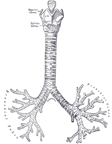
Anatomy of the trachea and bronchi (from Gray’s anatomy)
A. The ‘pump handle’ action of the ribs increases the transverse diameter of the chest
B. The diaphragm is responsible for abdominal breathing
C. Forced inspiration requires the use of rectus abdominis
D. Forced expiration requires the use of pectoralis minor
E. The ‘bucket handle’ action of the ribs is their upward and lateral movement
Show Answer
The ‘pump handle’ action of the ribs is their forward and upward movement increasing the antero-posterior diameter of the chest.
The ‘bucket handle’ action of the ribs is their upward and lateral movement increasing the transverse diameter of the chest.
Forced inspiration requires the use of the accessory muscles of respiration:
- Sternocleidomastoid
- Scalenes
- Pectoralis major
- Pectoralis minor
- Serratus anterior.
Forced expiration requires the use of:
- Rectus abdominis
- External and internal oblique
- Transversus abdominis
- Latissimus dorsi.
The diaphragm is responsible for abdominal breathing.
For thousands more anatomy tutorials and questions visit: www.anatomyprep.co.uk

Header image used on licence from Shutterstock.


Good job
So good
Soo good for learning and preparing exams
Very Useful and helpful
EXCELLENT. VERY EDUCATIVE QUESTIONS
good job
Very good
Good for revision, it really helped me