Test your knowledge of upper limb anatomy with these questions.
1. Which of the following muscles is LEAST likely to be affected in the presence of an ulnar nerve lesion?
A. Palmar interossei
B. Lateral two lumbricals
C. Dorsal interossei
D. Medial two lumbricals
E. Abductor digiti minimi
Show Answer
Answer: B. Lateral two lumbricals
The ulnar nerve innervates the following hand muscles:
- Palmar interossei
- Dorsal interossei
- Medial two lumbricals
- Abductor digiti minimi
The lateral two lumbricals are innervated by the median nerve and will not be affected by an ulnar nerve lesion.
2. Which SINGLE statement regarding the axillary artery is true?
A. It originates from the arch of the aorta
B. The axillary vein lies lateral to it
C. It provides blood supply to the humerus
D. It is divided into 3 parts by pectoralis major
E. After passing the lower margin of teres major it becomes the subclavian artery
Show Answer
Answer: C. It provides blood supply to the humerus The axillary artery is a continuation of the subclavian artery. It commences at the lateral border of the 1st rib and ends at the inferior border of teres major to become the brachial artery. It is divided into three parts by pectoralis minor:
- First part – medial to the upper border of pectoralis minor
- Second part – has pectoralis minor lying anterior to it
- Third part – extends from lower border of pectoralis minor to the inferior border of teres major
The axillary artery provides blood to the humerus via the anterior and posterior humeral circumflex arteries.
The axillary artery and its relations (from Gray’s Anatomy)
3. The rotator cuff will be affected by an injury to which of the following muscles?
A. Teres major
B. Subscapularis
C. Sternocleidomastoid
D. Pectoralis minor
E. Deltoid
Show Answer
Answer: B. Subscapularis
The rotator cuff muscles are a group of four muscles that originate from the scapula and attach to the humeral head. Acting together these muscles act to hold the humeral head in the glenoid fossa, stabilizing the shoulder joint.
The rotator cuff comprises the following four muscles:
- Supraspinatus
- Infraspinatus
- Subscapularis
- Teres minor
Anatomy of the shoulder showing teres minor, infraspinatus and supraspinatus (from Gray’s anatomy)
4. Which of the following best describes the sensory area supplied by the superficial branch of the radial nerve?
A. Part of the posterior aspect of the upper arm
B. A tapered strip of the middle portion of the posterior forearm
C. The lateral aspect of the anterior upper arm between the deltoid and the elbow
D. The posterior surface of the lateral three and a half digits and the associated areas of the palm
E. The palmer surface and fingertips of the lateral three and a half digits
Show Answer
Answer: D. The posterior surface of the lateral three and a half digits and the associated areas of the palm
The sensory function of the radial nerve is provided by its four main sensory branches:
- Inferior lateral cutaneous nerve of the arm – supplies the lateral aspect of the anterior upper arm between the deltoid and the elbow
- Posterior cutaneous nerve of the arm – supplies part of the posterior aspect of the upper arm
- The posterior cutaneous nerve of the forearm – supplies a tapered strip of the middle portion of the posterior forearm
The superficial branch of the radial nerve – supplies the posterior surface of the lateral 3 ½ digits and the associated areas of the palm
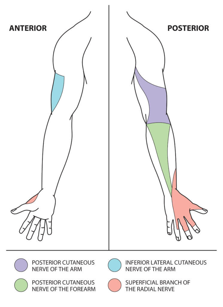
Image © Medical Exam Prep
5. Which of the following muscles is supplied the superficial branch of the ulnar nerve?
A. Flexor digiti minimi brevis
B. Dorsal interossei
C. Palmar interossei
D. Flexor digitorum profundus
E. Palmaris brevis
Show Answer
Answer: E. Palmaris brevis
The muscles supplied by the ulnar nerve and its two main motor branches are shown in the table below:
| Branch | Muscles supplied |
|---|---|
| Muscular branches of the ulnar nerve (in the forearm) | Flexor carpi ulnaris Flexor digitorum profundus (medial ½) |
| The deep branch of the ulnar nerve (in the hand) | Hypothenar muscles - Opponens digiti minimi - Abductor digiti minimi - Flexor digiti minimi brevis 3rd and 4th lumbricals Dorsal interossei Palmar interossei Adductor pollicis Flexor pollicis brevis (deep head) |
| The superficial branch of the ulnar nerve (in the hand) | Palmaris brevis |
For thousands more anatomy tutorials and questions visit: www.anatomyprep.co.uk
Header image used on licence from Shutterstock.


NICE , REALLY HELPED
Very nice….Thanks very much
🥰
I love this
thank you
I really love this, it’s great.
Really hard tho 😜 but thanks!
It was soo short :(( but the questions were really good! thanks ^-^
This was great. Please give more questions!
Great work i enjoyed it
Thanks for the explanations.
It will be nice to have more questions!
i like this work , great
Wow amazing
Amazing
This was indeed great