The ascending tracts are neural pathways located in the white matter that conduct afferent information from the peripheral nerves to the cerebral cortex.
There are usually three neurons in an ascending pathway:
- First-order neurons, in which the cell body is in the posterior root ganglion
- Second-order neurons, which decussate (cross to the opposite side) and ascend to a level higher up in the central nervous system
- Third-order neurons, which are located in the thalamus and pass to a sensory region of the cortex.
The main ascending tracts are:
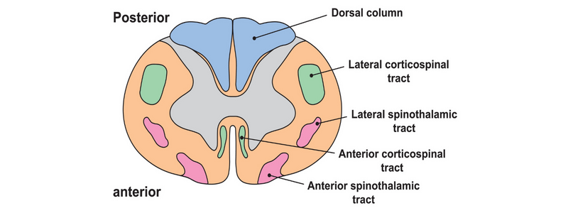
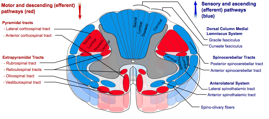
- The spinothalamic tracts (anterolateral system):
- The anterior spinothalamic tract (light touch and pressure pathway)
- The lateral spinothalamic tract (pain and temperature pathway)
- The spinocerebellar tracts:
- Anterior (ventral) spinocerebellar tract
- Posterior (dorsal) spinocerebellar tract
- The dorsal column-medial lemniscal pathway:
- The posterior white column: fasciculi gracilis and cuneatus (proprioception, fine touch, vibration to the postcentral gyrus)
Courtesy of Mikael Haggstrom CC BY-SA 3.0
The spinothalamic tracts
Both of the tracts in this system have three groups of neurons. The first-order neurons come from the peripheral sensory receptors. Once they enter the spinal cord, they ascend 1 or 2 vertebral levels via Lissauer’s tract, synapsing in the susbstantia gelatinosa, which is located at the edge of the dorsal horn.
The second-order neurons transmit information from the substantia gelatinosa to the thalamus. After synapsing with the first-order neurons, the fibres decussate in the spinal cord via the anterior white commissure and then form two distinct tracts:
- The anterior spinothalamic tract – conducts information about pressure and light (crude) touch
- The lateral spinothalamic tract – conducts information through the white matter of the spinal cord about pain and temperature
Third-order neurones transmit sensory signals from the thalamus to the primary sensory cortex on the same (ipsilateral) side of the brain. They originate in the ventral posterolateral nucleus of the thalamus, ascend through the internal capsule, and terminate in the sensory cortex.
The spinocerebellar tracts
These tracts transmit fibres from the muscles and terminate in the ipsilateral cerebellum. They carry information regarding proprioception. Whilst the other ascending tracts detect external stimuli from skin surface receptors, this tract detects stimuli from internal receptors regarding the length and tension of muscle fibres.
The origin of the proprioceptive information is within Golgi tendon organs and muscle spindles. These are first-order neurons, with their cell bodies in the dorsal root ganglion. They pass through the dorsal horn and form synapses with second-order neurons.
There are four individual pathways:
- Posterior (dorsal) spinocerebellar tract – carries proprioceptive information from the lower limbs to the cerebellum on the same (ipsilateral) side. Third-order neurones travel via the inferior cerebellar peduncle to reach the cerebellar cortex.
- Anterior (ventral) spinocerebellar tract – also transmits proprioceptive input from the lower limbs to the ipsilateral cerebellum. In this pathway, third-order neurones pass through the superior cerebellar peduncle to terminate in the cerebellar cortex.
- Rostral spinocerebellar tract – this carries proprioceptive information from the upper limbs to the ipsilateral cerebellum.
- Cuneocerebellar tract – this carries proprioceptive information from the upper limbs to the ipsilateral cerebellum.
The dorsal column-medial lemniscal pathway
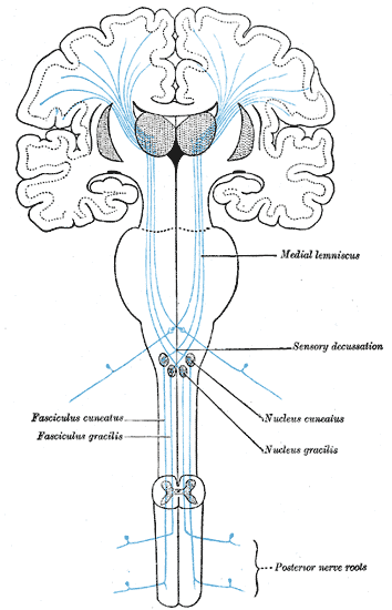
This pathway carries information regarding fine touch, two-point discrimination, vibration and proprioception from the skin and joints. The receptors involved include muscle spindles, Pacinian Corpuscles and hair follicle receptors.
The first-order neurons are located in the dorsal root ganglia, with their axons extending through to fasciculus gracilis and fasciculus cuneatus to the nuclei gracilis and cuneatus (in the lower medulla), where they synapse with the second-order neurons. These neurons then pass to the thalamus, where the third-order neurons synapse and send their axons to the postcentral gyrus.
Header image used on licence from Shutterstock
For thousands of anatomy tutorials and questions visit: www.anatomyprep.co.uk