The scalp is the area of the head that is bordered by the face at the front and the neck at the sides and back.

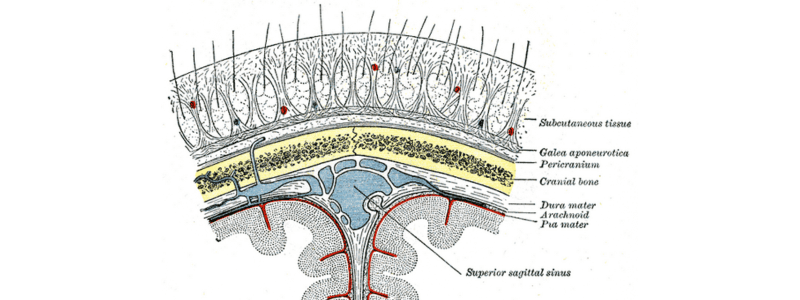
The layers of the scalp
It is classically described as having five layers with the helpful mnemonic ‘SCALP’:
- Skin
- Connective tissue
- Aponeurosis
- Loose connective tissue
- Pericranium
Skin: This layer contains numerous hair follicles and sebaceous glands. The hair follicles are often clustered together.
Connective tissue: This is a dense subcutaneous layer of fat and fibrous tissue that connects the skin to the epicranial aponeurosis and contains the nerves and vessels of the scalp. The blood vessels within this layer are highly adherent to the connective tissue.
Aponeurosis: This is a thin, tough layer that connects the frontalis muscle anteriorly to the occipitalis posteriorly.
Loose connective tissue: This layer is also known as the loose areolar connective tissue layer, and it separates the epicranial aponeurosis from the pericranium of the skull. It contains emissary veins that connect the veins of the scalp to the intracranial venous sinuses and the diploic veins. The connective tissue comprises various collagen bundles and allows the superficial layers of the scalp to be mobile in relation to the pericranium.
Pericranium: This is the periosteum of the skull bones, providing nutrients to them. At the suture lines, it becomes continuous with the endosteum.
Vasculature of the scalp
The scalp has an excellent blood supply from five pairs of arteries. Two of these are branches of the internal carotid artery, and three are branches of the external carotid artery.
The branches of the internal carotid artery supplying the scalp are:
- The supratrochlear artery accompanies the supratrochlear nerve and is a branch of the ophthalmic branch of the internal carotid artery. It supplies part of the forehead above the eye and anastomoses with the supraorbital artery and with the supratrochlear artery on the other side.
- The supraorbital artery is also a branch of the ophthalmic branch of the internal carotid artery. It accompanies the supraorbital nerve and supplies the lateral forehead and scalp.
The branches of the external carotid artery supplying the scalp are:
- The superficial temporal artery supplies a large part of the scalp via its frontal and parietal branches. These are mainly located in the frontal and temporal areas. It anastomoses with the supraorbital artery.
- The posterior auricular artery supplies the area of the scalp superior to and posterior to the auricle.
- The occipital artery lies posteriorly on the scalp and supplies much of this area.

The venous drainage of the scalp follows the arteries, with drainage occurring into the supratrochlear, supraorbital, superficial temporal, posterior auricular, and occipital veins. There is a connection between the scalp and dural venous sinuses via the emissary veins.
Lymphatic drainage of the scalp
Lymph vessels from the anterior half of the scalp drain to the parotid nodes, whilst those from the posterior half of the scalp drain to the occipital and posterior auricular nodes.
Innervation of the scalp
There are six main nerves that innervate the scalp. These are branches of either the trigeminal nerve or the cervical nerves.
The branches of the trigeminal nerve that innervate the scalp are:
- The supratrochlear nerve is a branch of the ophthalmic division of the trigeminal nerve, and it supplies the anteromedial forehead.
- The supraorbital nerve is also a branch of the ophthalmic division of the trigeminal nerve, and it supplies the scalp region between the anterolateral forehead and the vertex.
- The zygomaticotemporal nerve is a branch of the maxillary division of the trigeminal nerve and supplies the region of the temple.
- The auriculotemporal nerve is a branch of the mandibular division of the trigeminal nerve and supplies the skin supero-anteriorly to the auricle.
The branches of the cervical nerves that innervate the scalp are:
- The greater occipital nerve is a branch of the anterior rami of cervical nerves C2 and C3. It supplies the posterior aspect of the scalp up to the vertex.
- The lesser occipital nerve is also a branch of the anterior rami of cervical nerves C2 and C3. It supplies the area posterior to the auricle.

Scalp lacerations
The scalp has an excellent vascular supply, as outlined above. Lacerations to the scalp can cause profuse bleeding for several reasons:
- The blood vessels lie within the connective tissue layer of the scalp and are extremely adherent to this layer. This adherence prevents the normal vasoconstriction that occurs when a vessel is damaged.
- The arteries themselves form a large area of anastomoses, which contribute to the bleeding when damaged.
It is important to remember that most of the blood supply to the skull comes from the middle meningeal artery; therefore, if the blood supply to the scalp is interrupted, bone necrosis will not occur.
Images from Gray’s anatomy
For thousands of anatomy tutorials and questions visit: www.anatomyprep.co.uk


