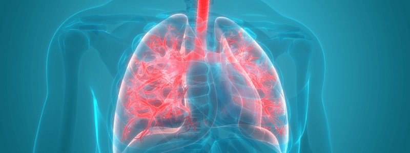
To ensure that enough oxygen is provided by ventilation to saturate the blood fully requires that ventilation and perfusion of the lungs are adequately matched. Ventilation (V) refers to the flow of air into and out of the alveoli, while perfusion (Q) refers to the flow of blood that reaches the alveoli via the capillaries.
The V/Q ratio
The ventilation/perfusion ratio (V/Q ratio) is a ratio used to the efficiency and adequacy of the matching of these two variables.
In an average 70 kg male:
- V is approximately 5 L/min
- Q is approximately 5 L/min
- The V/Q ratio is, therefore, ideally 1
Regional variation in the V/Q ratio
The V/Q ratio, however, varies in different regions of the lung partially because of the proximity of the heart to the region of the lung. Both ventilation and perfusion increase from the apex to the base of the lungs, perfusion, however, rises at a greater rate. This results in the following pattern:
- V/Q is highest at the apex of the lung (where it is approximately 3.3)
- V/Q is lowest at the base of the lung (where it is approximately 0.63)
- For this reason, the overall average value for V/Q is approximately 0.8
Gravity also plays a significant role in determining the V/Q ratio. The blood pressure at the base of the lungs is higher than the blood pressure at the apex because of the weight of the column of blood. This increased pressure results in distension of the pulmonary blood vessels at the base and an increase in blood flow and perfusion. Conversely, the blood flow at the apex is generally lower, which results in decreased perfusion.
Gravity also affects the intrapleural pressure, causing the intrapleural pressure to be increased at the base relative to the apex. This results in more compliant alveoli at the base and, as a result, ventilation is highest at the base of the lung.
Although these gravitational effects on perfusion and ventilation act to partially cancel each other out, ventilation is less significantly affected, further contributing to the pattern of the V/Q ratio being highest at the apex and lowest at the base. In young people, these gravitational effects are relatively minor and have little effect on lung function. In the elderly, however, the effects can be significant and may contribute to a low pO2.
Ventilation-perfusion mismatch and right-to-left shunts
Any mismatch between ventilation and perfusion will be evident in the V/Q ratio. If perfusion is normal, but ventilation is reduced, the V/Q ratio will be <1, whereas if ventilation is normal, but perfusion is reduced, the V/Q ratio will be >1.
If the alveoli were ventilated but not perfused at all, then the V/Q ratio would be infinity. A clinical example of this would be a pulmonary embolus that has blocked a pulmonary artery. Perfusion in the region supplied by that artery would be reduced to zero, rising the V/Q ratio to infinity. As no gas exchange is occurring in this area because of the lack of perfusion, this is referred to as alveolar dead space.
If the alveoli were perfused but not ventilated at all, then the V/Q ratio would be zero. A clinical example of this would be the inhalation of a foreign body that prevented all ventilation to an area of the lung. Ventilation in that region would be reduced to zero, lowering the V/Q ratio to zero. Because this area is being adequately perfused, but no oxygen is being transferred to the blood arriving there, a physiological right-to-left shunt has been created.
This should not be confused with an anatomical right-to-left shunt, where blood does not physically enter the lungs and instead moves directly from the right ventricle to the left ventricle. The end result of both types of right-to-left shunt is, however, the same, with arterial blood having a low pO2 and a high pCO2.
Ventilation-perfusion mismatch in clinical practice
In clinical practice, disease processes tend to cause changes in the V/Q ratio that vary throughout the lung. An example of this is chronic obstructive pulmonary disease (COPD), which causes destruction of the alveoli in the lung, resulting in large air spaces and capillary loss. The large air spaces result in poor ventilation, whereas the capillary loss results in inadeuquate perfusion.
The disease process is not uniform throughout the lungs, which means some areas of the lungs have a high V/Q ratio, whereas others have a low V/Q ratio. These V/Q mismatches play an important role in contributing to the hypoxia and hypercapnia seen in COPD.
Compensation for ventilation-perfusion mismatch
The body can compensate for V/Q mismatches and normalise the V/Q ratio as long as the mismatches are confined to restricted areas of the lung.
The two main mechanisms by which this can occur are:
- Hypoxic vasoconstriction: In the presence of a low V/Q ratio (i.e. areas of poor ventilation), hypoxic vasoconstriction can occur, which redirects incoming blood to the affected area to other parts of the lung. This decreases the perfusion of the hypoxic region, raising the V/Q ratio.
- Bronchoconstriction: In the presence of a high V/Q ratio (i.e. areas of poor perfusion), the bronchi constrict, increasing the resistance and decreasing the ventilation of the affected area that is not well perfused. This reduces the amount of alveolar dead space that occurs and lowers the V/Q ratio.
Header image used on licence from Shutterstock
Thank you to the joint editorial team of www.mrcemexamprep.net for this article.


Thank you for the explanation. Very educative.