
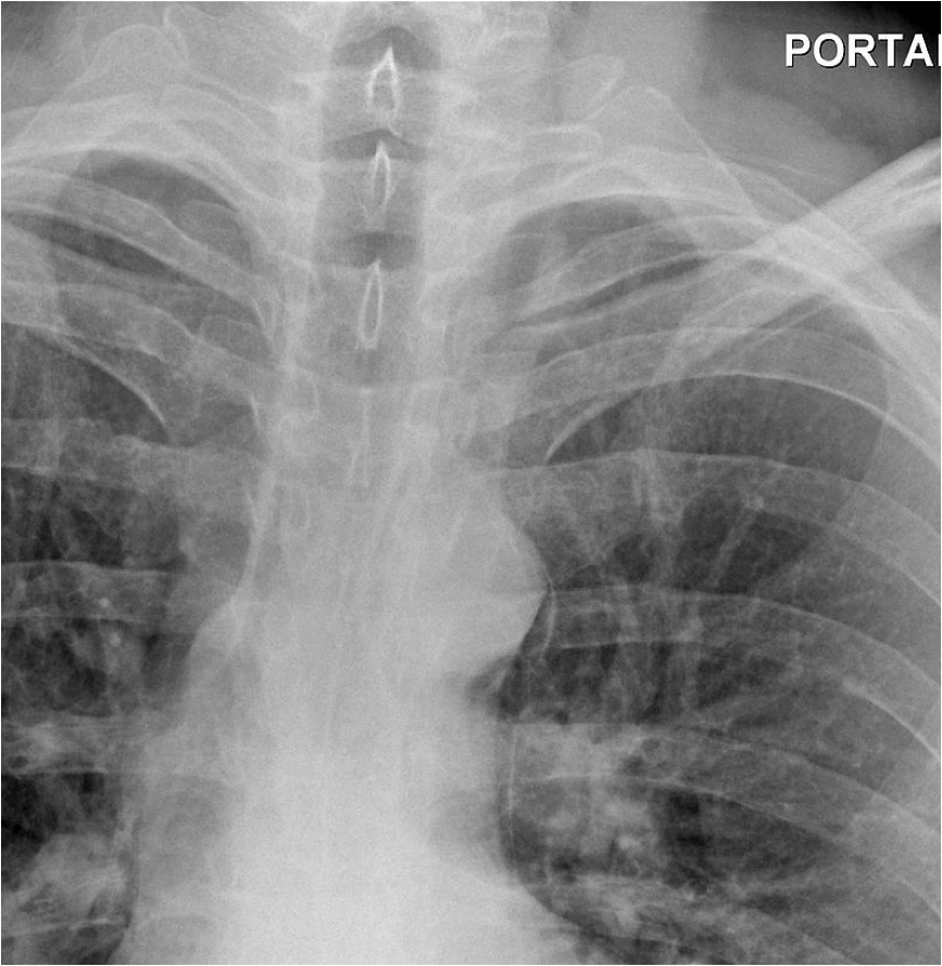
A 21-year-old medical student presents with severe retrosternal pain, pain on swallowing, and breathlessness. Over the weekend he went on a heavy drinking binge after finishing his exams and spent most of the previous evening vomiting and retching forcefully. You organise a chest X-ray, a portion of which is shown below:
1. What is the most likely diagnosis?
Show Answer
This patient has a diagnosis of Boerhaave’s syndrome.
Boerhaave’s syndrome is the spontaneous rupture of the oesophageal wall. The commonest cause is vigorous vomiting and retching. It has an association with excessive alcohol ingestion, when a dramatically raised pressure can develop in the oesophagus as a result of failure of cricopharyngeal relaxation.
Tears are usually vertically orientated and between 1 and 4 cm in length. Approximately 90% occur along the left posterolateral wall of the distal oesophagus.
The clinical features of Boerhaave’s syndrome include:
- Preceding history of vomiting
- Severe retrosternal and/or epigastric pain
- Back pain
- Failure of pain to be relived by opiate analgesia
- Breathlessness and cyanosis
- Subcutaneous emphysema (usually seen in neck)
- Hydrothorax
- Pneumothorax
- Pleural effusion
- Respiratory collapse
- Shock
2. Which supporting sign is present on the chest radiograph shown above?
Show Answer
Pneumomediastinum can be seen adjacent to the thoracic aorta and left common carotid artery:
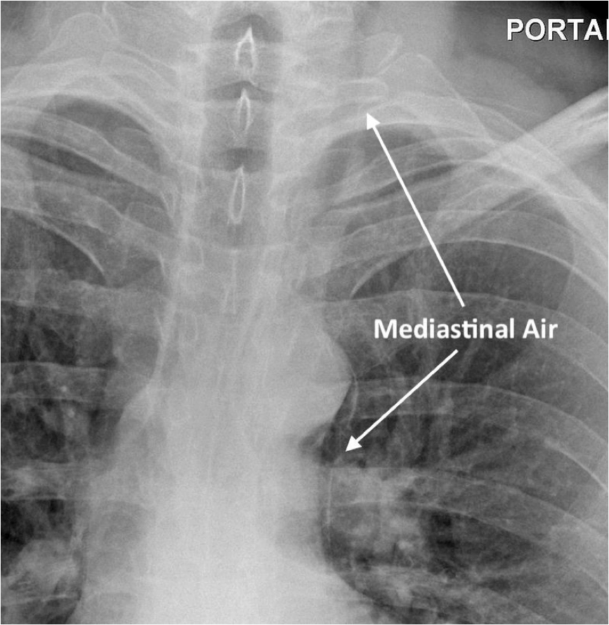
Radiograph demonstrating mediastinal air adjacent to the thoracic aorta and left common carotid artery, adapted from Wikipedia Courtesy of Jto410> CC BY-SA 3.0
3. Which other X-ray features that can be present with this condition?
Show Answer
Chest X-ray findings are non-specific, and the radiograph may be normal.
Changes that may be present on chest X-ray include:
- Pneumomediastinum
- Left-sided pleural effusion
- Pneumothorax
- Subcutaneous emphysema
- V sign of Naclerio (v-shaped air collection at left lower mediastinal border)
The diagnosis can be confirmed with a CT scan of the chest. Fluoroscopy can also be used, but has a 10% false negative rate.
4. What is the gold standard treatment for this condition?
Show Answer
The prognosis without treatment is poor and it is almost universally fatal. Mediastinal infection and sepsis occur rapidly and mortality is high if there is a delay in treatment.
Surgery is the gold standard for treatment, but the use of conservative methods, such as oesophageal stenting, have become more commonplace in recent years.
Header image used on licence from Shutterstock


Thanks, very interesting case
Well detailed explanation