Test your knowledge of the anatomy and physiology of the ECG with these questions.
1. Which of the following is the correct location of the sino-atrial (SA) node?
A. Right atrium
B. Left atrium
C. Right ventricle
D. Inter-atrial septum
E. Inter-ventricular septum
Show Answer
Answer: A. Right atrium
The sinoatrial node (SA node) is the pacemaker of the heart and is the point of origin of the electrical impulses that are propagated through the heart. The SA node is located in the right atrium and automatically generates an electrical impulse 60-100 times per minute under normal conditions.
These electrical impulses stimulate the atria to contract and then travel to the atrioventricular node (AV node), which is located in the inter-atrial septum. Here the impulse is briefly slowed before continuing down the conduction pathway to the bundle of His.
The bundle of His divides in the septum between the two ventricles into the left and right bundle branches, which are situated in the left and right ventricular muscle, respectively. Conduction then spreads out via specialised tissue within the ventricular walls known as Purkinje fibres.
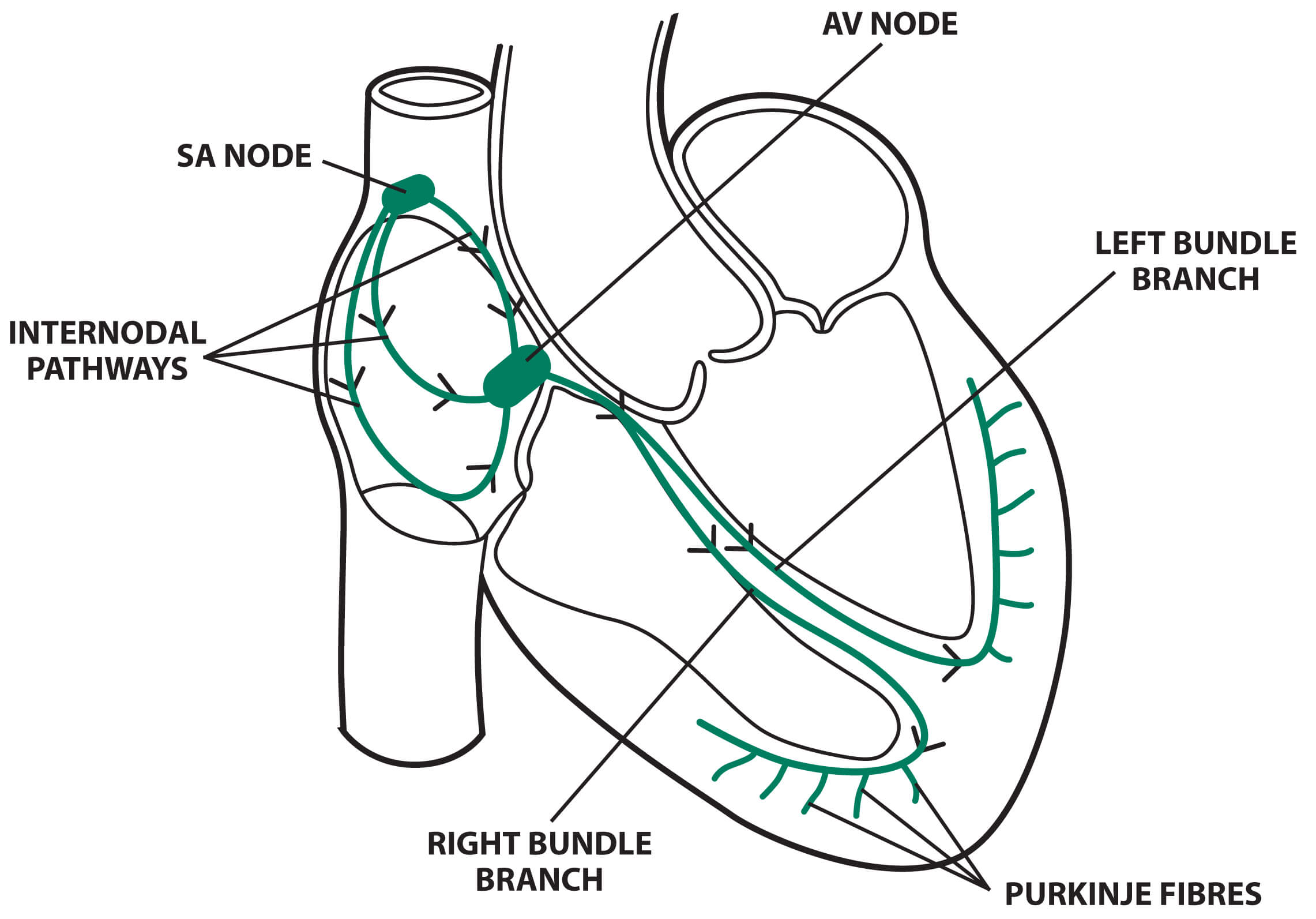
The cardiac conduction system © Medical Exam Prep
2. Which of the following is the correct location of the atrioventricular node (AV) node?
A. Right atrium
B. Left atrium
C. Right ventricle
D. Inter-atrial septum
E. Inter-ventricular septum
Show Answer
Answer: D. Inter-atrial septum
The atrioventricular node (AV node) is located in the inter-atrial septum.
3. At what rate does the atrioventricular node (AV) node fire under normal conditions?
A. 10-20 bpm
B. 20-40 bpm
C. 40-60 bpm
D. 60-80 bpm
E. 80-100 bpm
Show Answer
Answer: C. 40-60 bpm
The intrinsic rate of the AV node is 40 to 60 beats per minute (bpm).
4. Which SINGLE statement regarding the left coronary artery is true?
A. It is smaller than the right coronary artery
B. It supplies part of the bundle of His
C. It bifurcates into the anterior interventricular branch and the left marginal branch
D. It supplies the SA and AV nodes
E. It continues to the apex of the heart as the posterior descending artery
Show Answer
Answer B. It supplies part of the bundle of His
The left coronary artery arises from the aorta above the left cusp of the aortic valve. It supplies the left atrium, left ventricle, intraventricular septum and part of the left branch bundle of His. It is larger than the right coronary artery.
It passes between the left side of the pulmonary trunk and the left auricle and then bifurcates into the left anterior descending artery (LAD), which is also called the anterior interventricular branch, and the left circumflex artery (LCA). Sometimes an additional artery arises at the bifurcation called the ramus or intermediate artery.
The LAD follows the anterior interventricular groove towards the apex of the heart and then continues to the posterior surface of the heart to anastomose with the posterior interventricular branch.
The LCA follows the anterior interventricular groove to the left border of the heart and then proceeds to the posterior surface. It then gives rise to the left marginal branch, which follows the left border of the heart.
The right coronary artery arises above the right cusp of the aortic valve. It supplies the right atrium, the SA and AV nodes and the posterior part of the interventricular septum.
It runs along the right atrioventricular groove before branching. The right marginal artery arises first, and then the right coronary artery continues to the posterior surface of the heart, still running in the right atrioventricular groove. The posterior descending artery (PDA), which is also called the posterior interventricular artery, arises next and then follows the posterior interventricular groove towards the apex of the heart.
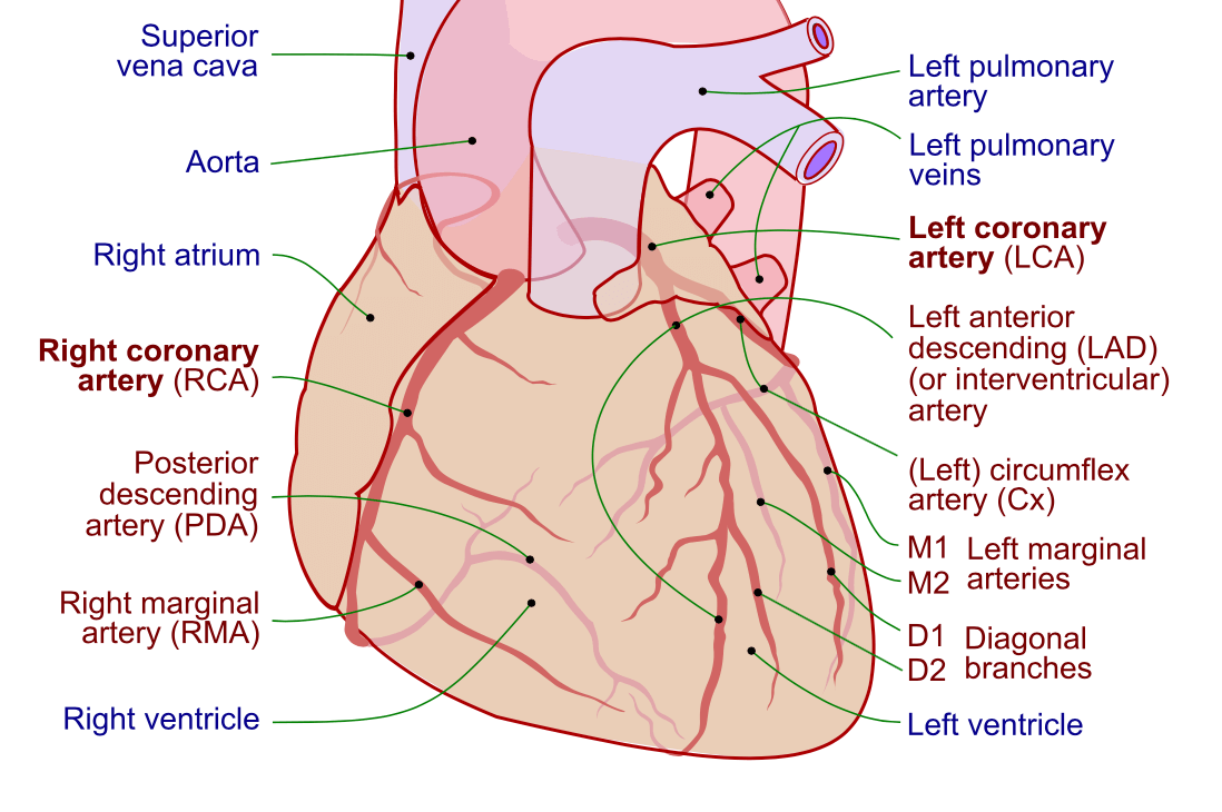
Image sourced from Wikipedia
Courtesy of Patrick J. Lynch CC BY-SA 3.0
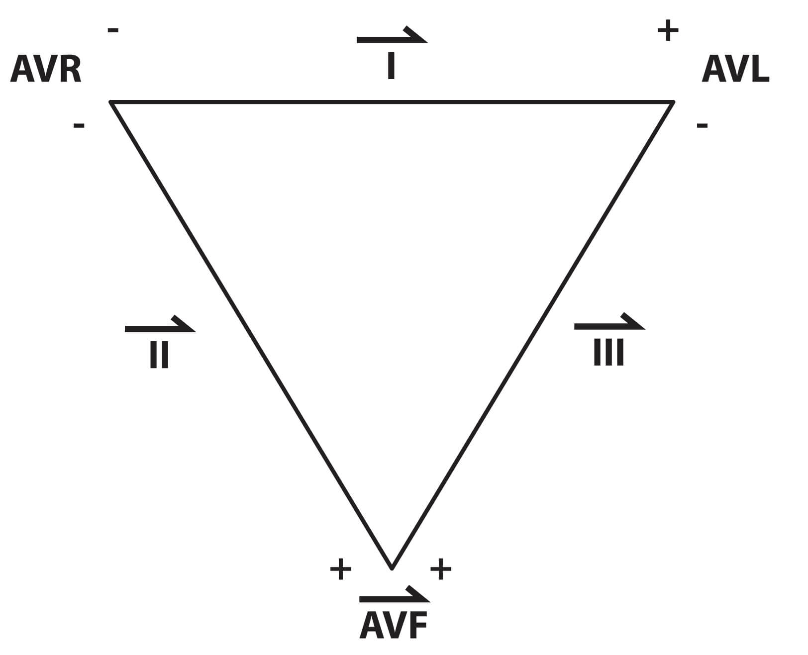
5. Which SINGLE statement regarding Einthoven’s triangle and the hexaxial system is true?
A. AVR, AVL and AVF are bipolar leads
B. The voltages created by AVR, AVL and AVF are large
C. Lead II gathers information between AVR and AVF
D. Lead III gathers information between AVR and AVL
E. Leads I, II and III each have one associated electrode
Show Answer
Answer: C. Lead II gathers information between AVR and AVF
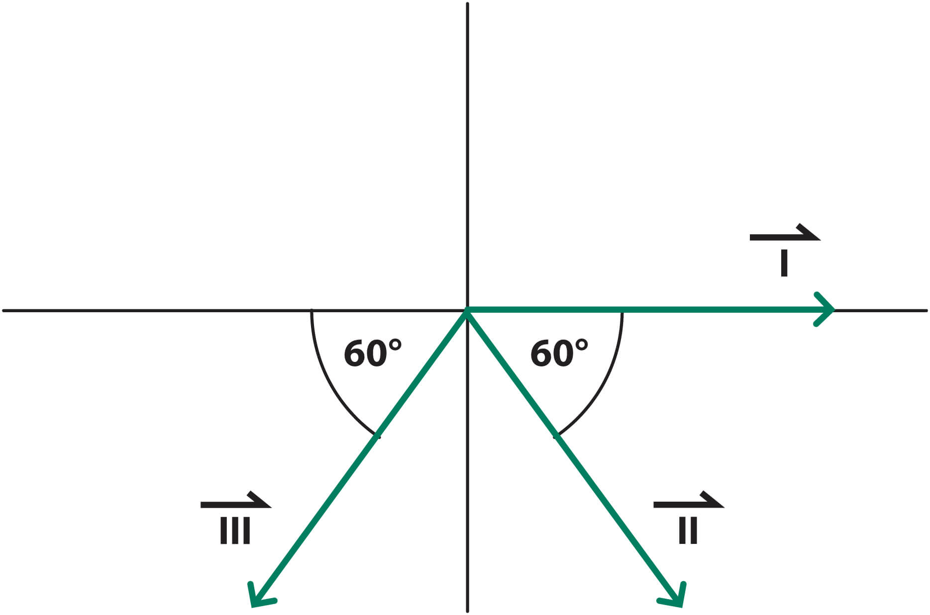
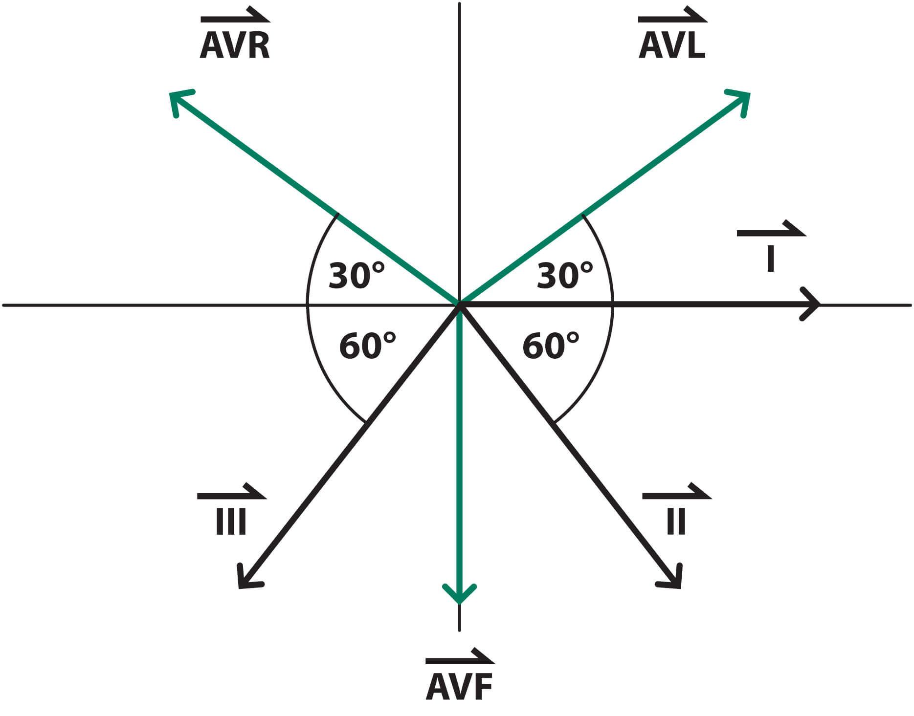
The limb leads are leads AVR, AVL, AVF, I, II and III. Each of these leads represents a measured voltage that also has a direction. By combining the magnitude of the measured voltage and the direction of the voltage, a vector is formed.
The limb leads AVR, AVL, AVF are also unipolar leads as they only have one associated electrode. The voltages of these electrodes are very small and have to be ‘augmented’ by built-in amplifiers. The ‘AV’ used in the terminology for these leads, therefore, stands for ‘Augmented Vector’. For these leads, the negative pole is once again the centre of the heart, and the three leads create a triangle with the heart in the middle. The vectors created are shown below:
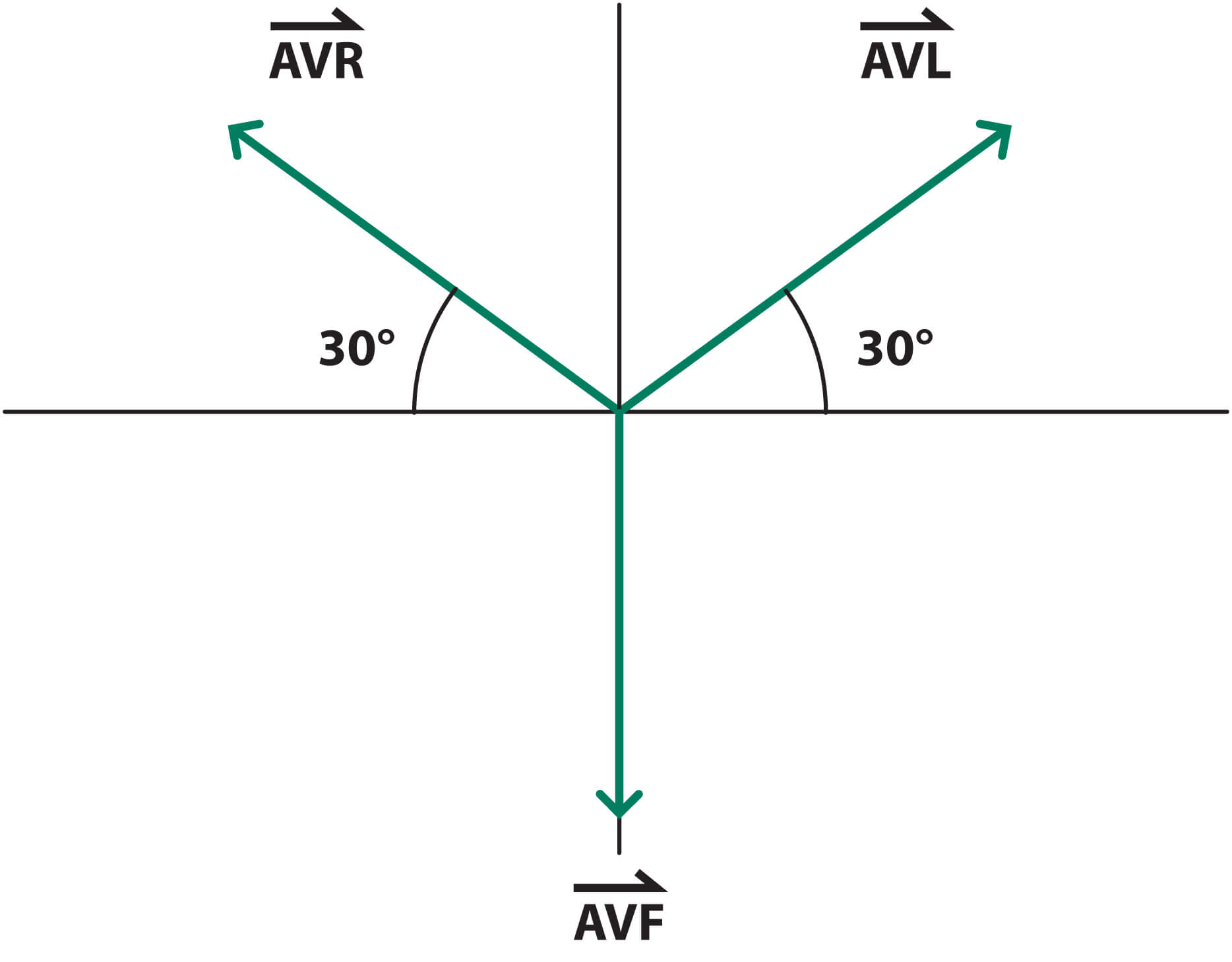
The limb leads I, II and III are called bipolar leads because they have two associated electrodes. AVR, AVL and AVF make up an equilateral triangle, known as ‘Einthoven’s Triangle’:
Information is gathered between these leads to create three more vectors:
- Lead I – information between AVR and AVL
- Lead II – information between AVR and AVF
- Lead III – information between AVL and AVF
The sum of these vectors is shown below:
Finally, by combining all 6 of these vectors together, we create the ‘hexaxial system’, which gives us a perspective of the view of all six of the limb leads.
All images © Medical Exam Prep
Header image used on licence from Shutterstock
Learn more about the ECG with our FREE ECG eBook ‘The Beginner’s Guide to the ECG’


Nice topic
very usefull
Excellent
Thank you for sharing such questions.
very useful revision